 Products Home
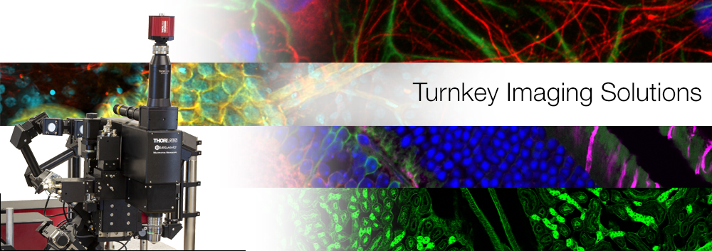

Purpose-Built Imaging Platforms
Thorlabs' microscopy systems combine the full benefits of a vertically integrated organization with decades of expertise to tailor virtually every component incorporated into its platforms. Our scientists and engineers draw from experience in all key disciplines to design, develop, manufacture, and support advanced imaging systems.
Our systems are designed from a clean sheet of paper to do the job intended from the beginning. Leveraging our extensive in-house design and fabrication capabilities allows us to minimize design trade-offs, develop targeted solutions to challenging problems, and customize each individual system to our customers' specifications. See below to view our imaging systems available.


Click to Enlarge
Two- and Three-Photon Imaging Configuration
Bergamo II Series Multiphoton Microscopes
Recognizing that there is a need for a microscope with a modular structure that can be configured to the experiment, rather than the other way around, we set out to develop the new reference standard in multiphoton microscopy. With a selection of rigid bodies, numerous auxiliary imaging modalities, detectors, motion control, and laser beam control, the permutations of the Bergamo II Series are endless. All configurations can be paired with the Tiberius® Ti:Sapphire Femtosecond Laser, which has an industry-leading tuning speed and is designed to enable fast sequential imaging in multiphoton microscopy applications.
For additional information, please see the full Bergamo II Series presentation.

Click to Enlarge
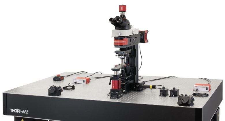
Veneto Inverted Microscope: Two-Photon + Widefield Configuration
Veneto Inverted Microscopes
Thorlabs’ Modular Inverted Microscopy Platform provides a turnkey solution for widefield, confocal, and multiphoton imaging. Inverted microscopes are powerful and versatile research tools that can accommodate a range of applications, such as fluorescence, in vivo, ex vivo, 3D, high-resolution, high-speed (video-rate), and live tissue imaging. This platform is available as a turnkey system that also offers users access to internal optomechanics and optics for DIY customization. We offer configurations that support widefield, darkfield, phase, Dodt, confocal, and multiphoton imaging.
For additional information, please see the full Veneto Inverted Microscopes presentation.
Click to Enlarge
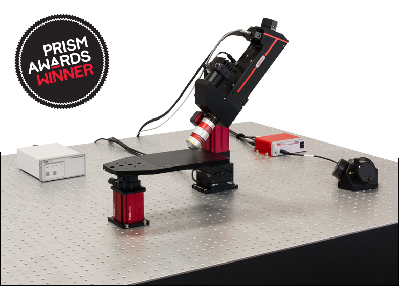
Prelude Microscope Shown with a TL10X-2P Objective Attached to a PFM450E Piezo Objective Scanner
Prelude Functional Imaging Microscope
Ushering in a new level of flexibility for functional imaging, Thorlabs has created a completely integrated two-photon microscopy system suitable for samples with demanding positioning requirements. The Prelude's compact fiber-coupled design eliminates the need for complex alignment procedures and provides a high degree of maneuverability. It includes a near-infrared 920 nm femtosecond pulsed laser and integrated filter set, making it ideally suited for two-photon fluorescence imaging of GFP. With a selection of long working distance objectives and a vibrationless remote focus, imaging with the Prelude can be done with no disturbance to your sample.
For additional information, please see the full Prelude presentation.

Mini2P Miniature Two-Photon Microscope
Thorlabs' Miniature Two-Photon Imaging System (Mini2P) is a fast, high-resolution miniscope designed for in vivo two-photon imaging of non-stationary specimens. The lightweight, head-mounted design allows for stable calcium imaging without impediment to the sample. Interchangeable objectives facilitate use with cranial windows, prisms, and GRIN lenses for imaging tissue at different depths and orientations. The Mini2P allows for hundreds of neurons to be monitored in a single FOV, while visualization of up to a thousand cells is achievable through multiplane imaging controlled through the built-in microtunable lens.
For additional information, please see the full Mini2P presentation.
Click to Enlarge
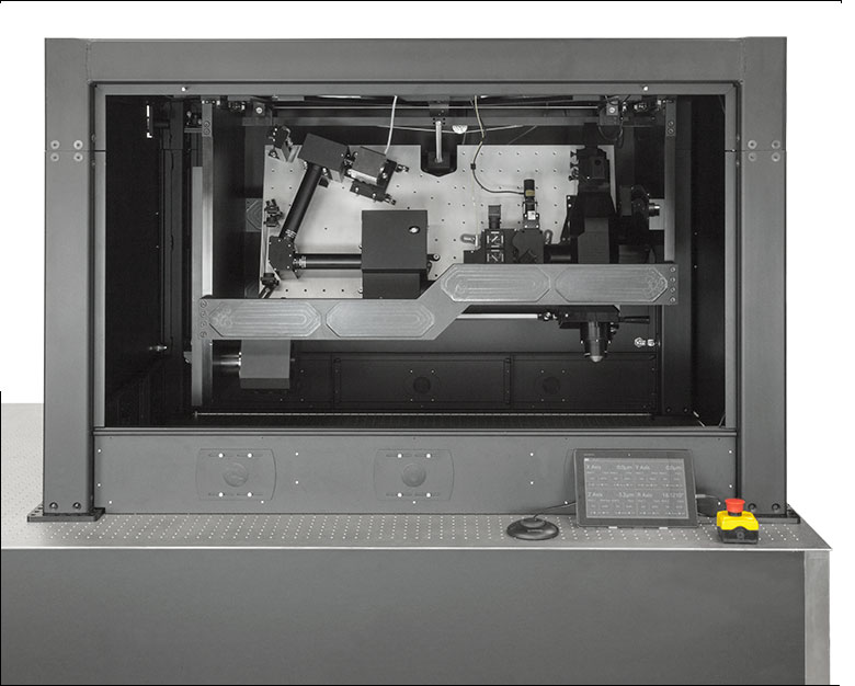
The Multiphoton Mesoscope Utilizes Technology Under License with HHMI's Janelia Research Campus
Multiphoton Mesoscope
To analyze large sections of the brain operating in concert, we developed a two-photon mesoscope for rapid functional imaging over a large 5 mm x 5 mm field of view (FOV). Scan over the entire FOV or rapidly image multiple spatially separated regions. The mesoscope enables in vivo awake-specimen neural studies at sub-cellular resolution with near-video imaging frame rates.
For additional information, please see the full Multiphoton Mesoscope presentation.

Click to Enlarge
Veneto Inverted Microscope: Confocal + Widefield Configuration
Confocal Microscopy Systems & Components
Thorlabs' Confocal Microscopy offerings include complete systems for laser-scanned fluorescence or reflected light microscopy and an assortment of component products for DIY construction or custom applications. Our systems are designed to be compatible with our Cerna modular microscope components, allowing them to be easily upgraded for additional functionality.
To view all of our confocal options, please see the Confocal Microscopy presentation.
Click to Enlarge
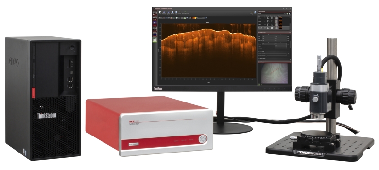
Ganymede™ Spectral Domain OCT Imaging System
Optical Coherence Tomography (OCT) Imaging Systems
Thorlabs offers a range of OCT imaging systems for various applications, including in vivo, vascular, ophthalmologic, and other biomedical imaging, as well as non-destructive testing (NDT), industrial inspection, and quality control. Our modular approach to OCT design provides a high degree of flexibility for optimizing your system to meet the unique needs of your application. Whether you are interested in a preconfigured system, or you prefer to select each component individually, please contact us. We are happy to assist you as you explore available options.
For additional information on our OCT imaging systems, please click here.
Click to Enlarge
Cerna DIY Microscope Configured for Widefield Imaging
Cerna Modular Microscopy Platform
The Cerna Series is Thorlabs' easily adjustable, DIY microscopy platform for diverse experimental requirements, including cellular and developmental biology, electrophysiology, optogenetics, and machine inspection. Its versatile, expandable, and accessible design is fully compatible with Thorlabs-manufactured and user-supplied accessories. Cerna microscopes are available preconfigured or can be custom built using our diverse selection of microscope components. Preconfigured Cerna-based systems are also available for simple imaging processes.
For additional information, please see the full Cerna Series presentation.
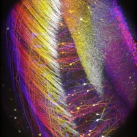
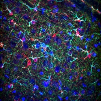
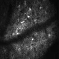
Images Above From Top Left to Bottom Right:
- Two-photon image of neurons expressing Thy1-YFP in a cleared region of a mouse hippocampus. (Courtesy of the 2017 Imaging Structure and Function in the Nervous System Course at Cold Spring Harbor Laboratory, Cold Spring Harbor, NY.)
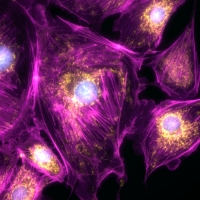
- Wild-type mouse brain section tagged with DAPI (405 nm), Alexa 488 anti-S100B, Alexa 555 anti-Neurofilament, and Alexa 633 anti-GFAP. (Sample Courtesy of Lynne Holtzclaw, NIH/NICHD/MIC.)
- Z-stack of a GFP-labeled mouse brain section taken with a Prelude Functional Imaging Microscope. Neurons are color-coded by depth.
- Two-photon calcium activity of a mouse medial entorhinal cortex imaged with a Mini2P. (Taken from Large-scale two-photon calcium imaging in freely moving mice.)
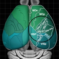
- Illustration depicting Mesoscope's ability to capture multiple brain regions simultaneously. The black circle represents a Ø5 mm FOV. (Taken from A Large Field of View Two-Photon Mesoscope with Subcellular Resolution for In Vivo Imaging and used here under a Creative Commons Attribution license.)
- Stitched image of a complete Drosophila fly head, using a 40X objective on a Thorlabs Confocal System.
- Sectioned 3D Image of a Zebrafish taken with a Thorlabs Telesto® OCT System
- Image of bovine pulmonary artery endothelial cells, acquired using a Cerna Microscope. (Courtesy of the Lab of Dr. Peter Stys, University of Calgary.)
Thorlabs recognizes that each imaging application has unique requirements.
If you have any feedback, questions, or need a quotation, please contact ImagingSales@thorlabs.com or call (703) 651-1700.
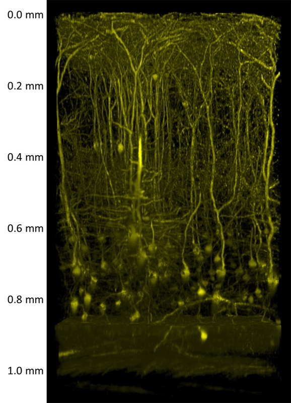
In Vivo Deep Tissue ImagingDeep tissue imaging is a hallmark of multiphoton microscopy. The Bergamo® II Series' ultrasensitive detector module utilizes full-field-of-view, non-descanned GaAsP PMT detectors to maximize the collection of photons scattered deep within a sample. This efficient design allows morphological features deep within a sample to be reconstructed without having to excise tissue. The image to the left shows an XZ projection of the primary somatosensory cortex in an 8-week-old mouse expressing thy1-YFP (H line), with a sample depth in excess of 1.0 mm. The excitation wavelength was 960 nm and the data was taken with a Nikon Apo LWD 25X objective with NA 1.10. The data set is courtesy of Dr. Hajime Hirase and Katsuya Ozawa, RIKEN Brain Science Institute, Wako, Japan. |
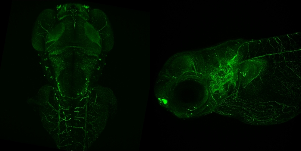
Developmental BiologyMaximum projection (dorsal and lateral, respectively) of zebrafish embryos, 48 hrs post-fertilization, showing developing neurons. Neurons are labeled with Alexa 488 against acetylated tubulin. These images were taken using Thorlabs' confocal systems. The fluorophore was excited using a 488 nm laser and imaged with an Olympus 20X 1.0 NA water dipping lens. The data set is courtesy of Aminah Giousoh and Dr. Robert Bryson-Richardson, School of Biological Sciences, Monash University, Victoria, Australia. |
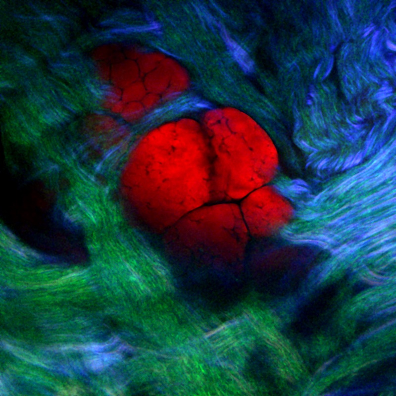 Click to Enlarge Merged Image Structural Insight with Nonlinear ImagingAlthough fluorescent proteins can overcome the limitations of thick or in vivo samples, where it is not always easy for fluorophores to penetrate, their use is not always desired. Nonlinear microscopy, of which two-photon fluorescence is a part, offers several complementary contrast mechanisms for visualizing structure. Coherent Anti-Stokes Raman Scattering (CARS), Second Harmonic Generation (two-photon fluorescence), and Third Harmonic Generation (three-photon fluorescence) are nonlinear imaging techniques that offer contrast that is the direct result of the interaction of the laser with the structure of interest. Under specific conditions, this allows the generation of an observable signal without the need for fluorophores. The figure to the right shows an image made by combining CARS, SHG, and Sum Frequency Generation (SFG) signals, while the figures below shows the individual signals from each of these imaging modalities. These images were taken using a Thorlabs multiphoton microscope. 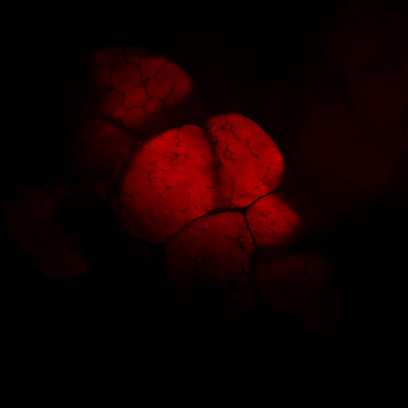 Click to Enlarge Epi-CARS 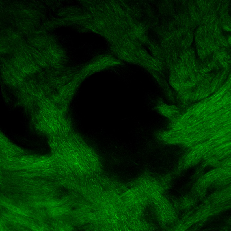 Click to Enlarge Epi-SHG 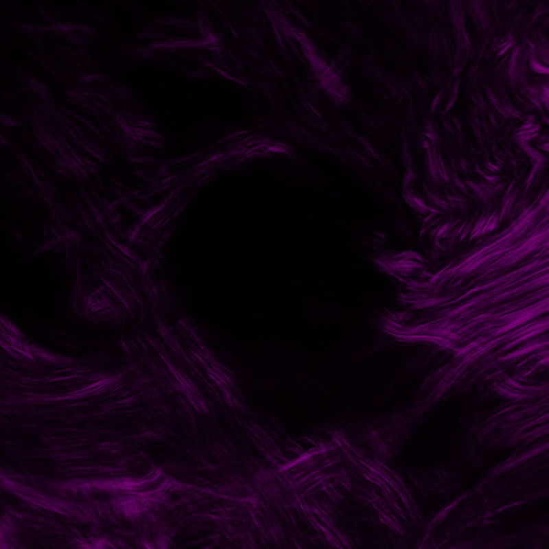 Click to Enlarge Forward SFG 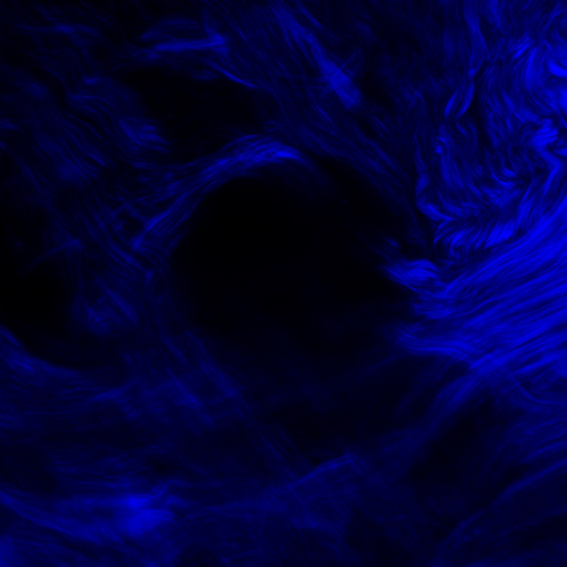 Click to Enlarge Forward SHG |
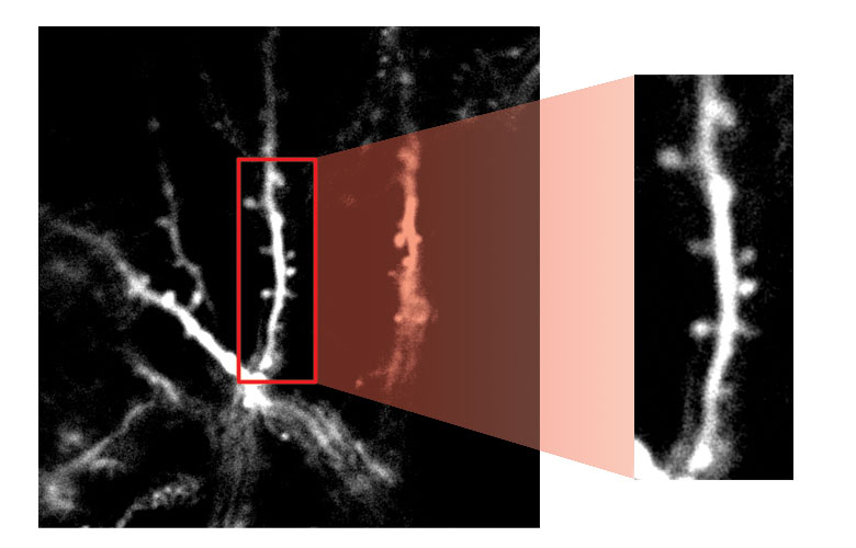 Click to Enlarge Dendritic Spines Expressing tdTomato High-Resolution ImagingExpanded view of dendritic spines from neurons in the mouse visual cortex expressing tdTomato. In this image, taken with a Thorlabs multiphoton microscope, the excitation wavelength was 1040 nm, and an Olympus 60X objective with NA 1.0 was used. This image is courtesy of Dr. Tobias Rose, Max Planck Institute for Neurobiology, Martinsreid, Germany. |
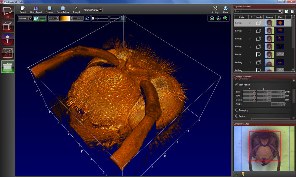 Click to Enlarge Image of a Wasp Cadaver in Volume Rendering Mode Deep 3D Volumetric ImagingAn image of a wasp cadaver. The optical video image of the sample can be seen in the bottom right corner. This image was taken with Thorlabs' Telesto® Series Spectral Domain OCT System. The excitation wavelength was 1300 nm. |
Thorlabs recognizes that each imaging application has unique requirements.
If you have any feedback, questions, or need a quotation, please contact ImagingSales@thorlabs.com or call (703) 651-1700.
Deep Insights
In Vivo Functional Neural Imaging of Large VolumesLarge FOV imaging at high spatiotemporal resolution is a necessary requirement for correlating neural activity of large brain volumes. This movie demonstrates the firing of neurons expressing GCaMP6f in behaving mice running on a spherical treadmill. The low magnification movie shows activity across multiple brain areas, including the somatosensory, parietal, and motor areas, at 4.3 Hz across a 6.3 mm x 5.4 mm FOV. The four non-contiguous regions were imaged at 9.6 Hz with 600 μm x 600 μm FOV, and fluorescence signals from individual neurons were sampled to cross-correlate their activity. In these image sets, taken with the Multiphoton Mesoscope, the excitation wavelength was 970 nm. The data set is courtesy of Nicholas James Sofroniew, Daniel Flickinger, Jonathan King, and Karel Svoboda; Janelia Research Campus and Vidrio Technologies, Virginia, USA (video taken from https://elifesciences.org/content/5/e14472/article-data and used here under a Creative Commons Attribution license). |
Intravital ImagingThe ability to image regions within an organ of a living animal (and not just very thin tissue, like mesentery tissue) represents a major advancement in intravital microscopy. In this movie, blood perfusion can be monitored. Blood is labeled with a tail vein injection of FITC-Dextran containing red fluorescent microspheres. The microspheres can be tracked to indicate blood velocity. Red blood cells are also visible as dark shadows within the green vessels. In these images, taken with a Thorlabs multiphoton microscope, the excitation wavelength was 780 nm and an Olympus XLUMPLFLN 20X objective with NA 1.0 was used. The preparation is courtesy of Dr. Simon Rhodes, IUPUI School of Medicine, Indianapolis, Indiana. |
Deep Brain ImagingDeep tissue imaging is a hallmark of multiphoton microscopy. The Bergamo® II Series' ultrasensitive detector module utilizes full-field-of-view, non-descanned GaAsP PMT detectors to maximize the collection of photons scattered deep within a sample. This efficient design allows morphological features deep within a sample to be reconstructed without having to excise tissue. The images shown above represent an XZ projection of the primary somatosensory cortex in an 8-week-old mouse expressing thy1-YFP (H line), with a sample depth in excess of 1.0 mm. The excitation wavelength was 960 nm and the data was taken with a Nikon Apo LWD 25X objective with NA 1.10. The data set is courtesy of Dr. Hajime Hirase and Katsuya Ozawa, RIKEN Brain Science Institute, Wako, Japan. |
Confocal Reflectance MicroscopyNot all samples can be labeled with an exogenous contrast agent. Confocal reflectance microscopy allows for optical sectioning imaging by observing reflected laser light. This movie represents a Z-stack of a peachworm. These images were taken with a Thorlabs confocal microscope. The excitation wavelength was 488 nm and the data was taken with an Olympus UPlanFl 20X objective with NA 0.50. |
Improved Image Contrast with Fast TuningWith an industry-leading tuning speed of up to 200 nm in <50 ms, the Tiberius® Ti:Sapphire Femtosecond Laser is ideal for fast sequential imaging. The Tiberius' fast-tuning capability provides high-contrast images when used in multi-color, multiphoton microscopy applications. Quickly switching between two optimized excitation wavelengths has several benefits over single-wavelength excitation. These include the much higher image contrast provided by fast switching and being able to maximize fluorescence at lower excitation powers, which reduces the risk of photobleaching. This video shows the real-time flashing between red and green fluorescence excited by the Tiberius Ti:Sapphire Laser's high-speed wavelength switching. Both channels were collected at an imaging rate of 7 fps with a resolution of 512 x 512 pixels. If you experience adverse effects from visual stimuli including flashing lights, please watch this version played at 1/16th the imaging rate as an alternative. This immunofluorescence sample was prepared by Lynne Holtzclaw of the NICHD Microscopy and Imaging Core Facility, a part of the National Institutes of Health (NIH) in Bethesda, MD. |
Thorlabs recognizes that each imaging application has unique requirements.
If you have any feedback, questions, or need a quotation, please contact ImagingSales@thorlabs.com or call (703) 651-1700.


Click to Enlarge
China Demo Room
Try Our Microscopes In Person or Virtually
Thorlabs' sales engineers and field service staff are based out of nine offices across four continents. We look forward to helping you determine the best imaging system to meet your specific experimental needs. Our customers are attempting to solve biology's most important problems; these endeavors require matching systems that drive industry standards for ease of use, reliability, and raw capability.
Thorlabs' worldwide network allows us to operate demo rooms in a number of locations where you can see our systems in action. We welcome the opportunity to work with you in person or virtually. A demo can be scheduled at any of our showrooms or virtually by contacting ImagingSales@thorlabs.com.
Customer Support Sites
(Click Each Location for More Details)
Newton, New Jersey, USA
Thorlabs Headquarters
43 Sparta Avenue
Newton, NJ 07860
Customer Support
- Phone: (973) 300-3000
- E-mail: techsupport@thorlabs.com
Ely, United Kingdom

Thorlabs Ltd.
1 Saint Thomas Place, Ely
Ely CB7 4EX
Customer Support
- Phone: +44 (0)1353-654440
- E-mail: techsupport.uk@thorlabs.com
Bergkirchen, Germany
Thorlabs GmbH
Münchner Weg 1
85232 Bergkirchen
Customer Support
- Phone: +49 (0) 8131-5956-0
- E-mail: europe@thorlabs.com
Le Mesnil-le-Roi, France
Thorlabs SAS
14 rue Gambetta
78600 Le Mesnil-le-Roi
Customer Support
- Phone: +33 (0) 970 440 844
- E-mail: techsupport.fr@thorlabs.com
São Carlos, SP, Brazil
Thorlabs Vendas de Fotônicos Ltda.
Rua Rosalino Bellini, 175
Jardim Santa Paula
São Carlos, SP, 13564-050
Customer Support
- Phone: +55-16-3413 7062
- E-mail: brasil@thorlabs.com
Demo Rooms and Customer Support Sites
(Click Each Location for More Details)
Sterling, Virginia, USA
Thorlabs Imaging Systems HQ
108 Powers Court
Sterling, VA 20166
Customer Support
- Phone: (703) 651-1700
- E-mail: ImagingTechSupport@thorlabs.com
Demo Rooms
- Bergamo® II Series Multiphoton Microscopes
- Veneto® Inverted Microscopes
- Four-Channel Cerna®-Based Confocal Microscopes
- Cerna Birefringence Imaging Microscopes
- Multiphoton Mesoscope
- OCT Systems: Telesto® and Ganymede™
Lübeck, Germany
Thorlabs GmbH
Maria-Goeppert-Straße 9
23562 Lübeck
Customer Support
- Phone: +49 (0) 8131-5956-40840
- Email: oct@thorlabs.com
Demo Rooms
- Ganymede™ Series SD-OCT Systems
- Telesto® Series SD-OCT Systems
- Telesto® Series PS-OCT Systems
- Atria® Series SS-OCT Systems
- Vega™ Series SS-OCT Systems
Nerima-ku, Tokyo, Japan
Thorlabs Japan, Inc.
3-6-3 Kitamachi
Nerima-ku, Tokyo 179-0081
Customer Support
- Phone: +81-3-6915-7701
- Email: sales@thorlabs.jp
Demo Rooms
- Four-Channel Cerna®-Based Confocal Systems
- Cerna® Modular Brightfield Microscopes
- OCT Systems: Ganymede™
Shanghai, China
Thorlabs China
Room A101, No. 100, Lane 2891, South Qilianshan Road
Shanghai 200331
Customer Support
- Phone: +86 (0)21-60561122
- Email: techsupport-cn@thorlabs.com
Demo Rooms
- Bergamo® II Series Multiphoton Microscopes
- Cerna Birefringence Imaging Microscopes
- OCT Systems: Telesto® and Ganymede™
Brochures
The buttons below link to PDFs of printable materials for Thorlabs imaging products and systems.


| Posted Comments: | |
Lingjie Kong
(posted 2024-04-18 18:53:17.253) Hi there,
We recently developed the random-access wide-field mesoscopy, which was published on Nature Photonics on April 17th 2024, at https://www.nature.com/articles/s41566-024-01422-1
I wonder if you are interested in getting it commercialized?
Feel free to contact me. Thanks!
Best,
Lingjie cdolbashian
(posted 2024-04-26 02:40:22.0) Thank you for reaching out to us Lingjie. We have contacted you directly to discuss your publication. |