 Products Home / Imaging Systems / Cerna® Modular Microscopy Platform / Cerna Preconfigured Microscopes / Cerna® Microscope with Epi-Illumination and Trans-Illumination Modules
Cerna® Microscope with Epi-Illumination and Trans-Illumination Modules
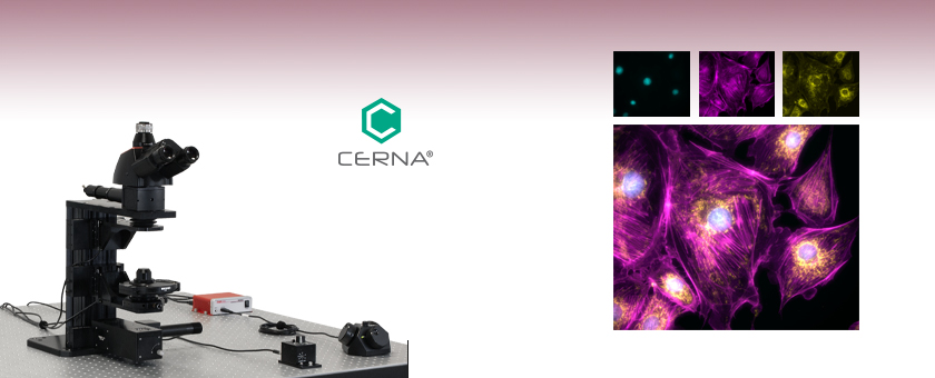
- Equipped with Single-Cube Epi-Illuminator and
Trans-Illumination Modules - Ready to Accept Objectives, Cameras, Filters,
and Illumination Sources
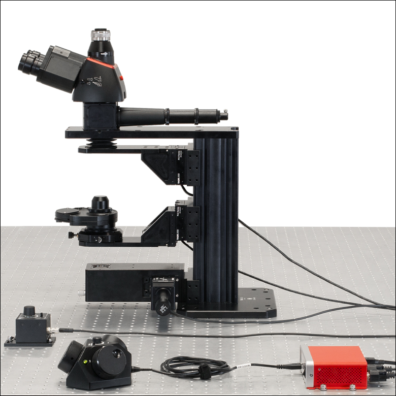
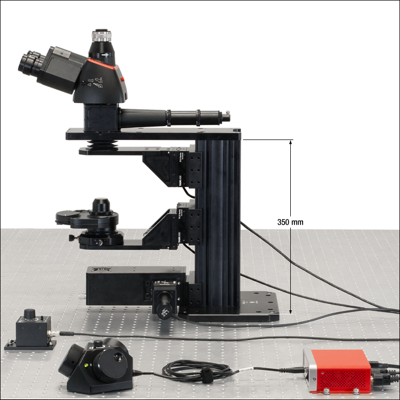
Cerna® Microscope Kit 3
(Optical Table Not Included)
Image of bovine pulmonary artery endothelial (BPAE) cells acquired using a Cerna Microscope with the Chrolis™ LED Source and CS505MU Camera. (Courtesy of the Lab of Dr. Peter Stys, University of Calgary.)
Nuclei:
DAPI
(365 nm)
Actin:
AF 488
(475 nm)
Mitochrondia:
MitoTracker™
Red (565 nm)


Please Wait
Features
- Single-Cube Epi-Illuminator and Transmitted Illumination Modules
- Epi-Illuminator Module Compatible with Thorlabs' Mounted LEDs, Chrolis™ High-Power LED Sources, and Ø3 mm Liquid Light Guides
- Transmitted Light Module Accepts LEDs for Visible and/or IR Light
- Accepts C-Mount Cameras from Thorlabs and Most Major Manufacturers
- Motorized Focus Control of Objective
- Trinoculars with 10X Eye Pieces
- Modular Design Allows User to Modify the Microscope's Optical Path
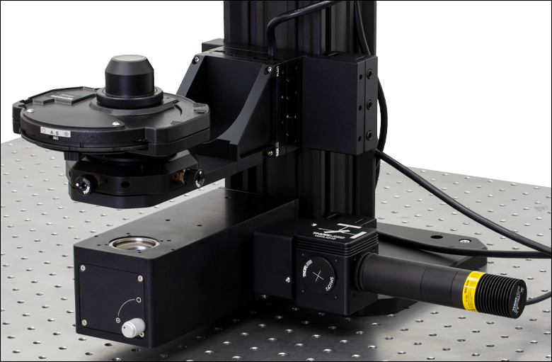
This Cerna® microscope configuration provides an optical path that is ideal for experiments requiring either epi-fluorescence, reflected light, or brightfield imaging. The epi-illumination module accepts a variety of LEDs and lamps equipped with a Ø3 mm liquid light guide, while the brightfield illumination module includes Thorlabs' Visible Illumination Kit. A motorized objective holder on the microscope body provides 1" of vertical focusing adjustment for the objective.
Trinoculars with a camera port support real-time viewing of the sample directly through the eyepieces. The C-mount-threaded camera port is compatible with most industry-standard cameras, which can be used to view the image on a computer screen in real time or to capture images to analyze later. Motorized objective and condenser focusing modules, each with 1" of travel, provide fine-tuned positioning along the optical axis.
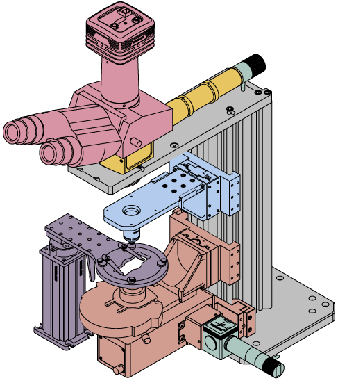
Unlike competing microscopes with similar capabilities, the Cerna platform's modularity lets the user quickly install and remove the microscope modules as needed for each experiment, providing a high degree of access and control. In vitro samples can be studied by positioning sample stages below the objective using fixed arms that can be attached directly to the microscope or rigid stands. To free room underneath the objective for large sample holding apparatuses, the brightfield module can be removed, providing a path for in vivo studies.
To address a wide range of experimental parameters, Thorlabs offers eight Cerna microscope configurations, which are summarized in Table 1.2. In addition, we can work with you to configure a microscope that meets your unique needs. To contact our team, please e-mail ImagingSales@thorlabs.com. We also offer Cerna components individually for custom modifications.
| Table 1.2 Cerna Microscope Kits | ||||||||
|---|---|---|---|---|---|---|---|---|
| Kit 1 | Kit 2 | Kit 3 | Kit 4 | Kit 5 | Kit 6 | Kit 7 | Kit 8 | |
| Objective Holder | Single | Single | Single | Dual | Dual | Dual | Dual | Dual |
| Epi-Illumination | 1 Cube | Up to 6 Filter Sets | 1 Cube | Up to 6 Filter Sets | Up to 6 Filter Sets | Up to 6 Filter Sets | Up to 6 Filter Sets | Up to 6 Filter Sets |
| Trans-Illumination | - | - | Brightfield (Visible) |
Brightfield (Visible) |
Dodt Contrast and Brightfield (Visible) |
Dodt Contrast and Brightfield (Visible and NIR) |
DIC Imaging and Brightfield (Visible and NIR) |
DIC Imaging and Brightfield (Visible and NIR) |
| XY Motion | - | - | - | - | - | Microscope Translator |
N/A | Translating Platform |
Cerna® Microscope Kit 3
This Cerna microscope kit was designed from our line of modular components to provide several convenient features for imaging, highlighted below. We also offer a selection of microscope objectives, cameras, and illumination modules that can be used to complement this microscope configuration and customize it to your experiment. Details can be found on the Microscope Add-Ons tab. The Kit Components tab details the components used in this microscope configuration, as well as a link to each component's webpage, where additional information (such as mechanical drawings) is available.
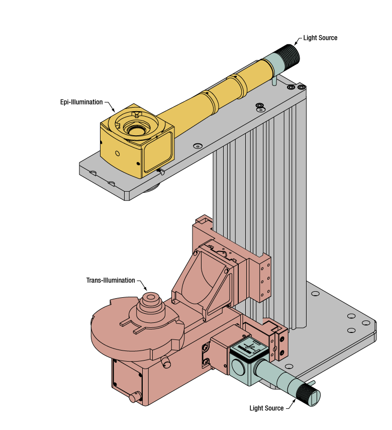
Epi-Illumination
Add-Ons: Epi-Illumination
Features
- Single-Cube Epi-Illuminator Module (Filter Cubes and Sets Sold Separately)
- Accepts Thorlabs' Mounted LEDs, Chrolis™ High-Power LED Sources, or Other Sources that Use Ø3 mm Liquid Light Guides
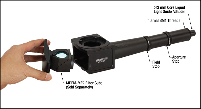
The epi-illumination module couples light emitted by the illumination source into the imaging path, through the objective, and onto the sample; it also allows epi-fluorescence generated by the sample to pass through the module to the eyepieces and camera. This epi-illuminator accepts one filter cube, making it suitable for several imaging modalities that require a single filter set. By installing a dichroic mirror and two emission filters, fluorescence imaging of a single fluorophore is possible. This filter set can be replaced with a 50:50 beamsplitter and two polarizers to create a reflected light imaging microscope. Alternatively, a multiband filter set combined with illumination from a multi-wavelength LED source allows the microscope to image samples with multiple fiducial markers. For more details about epi-illumination options offered by Thorlabs, please see the full web presentation.
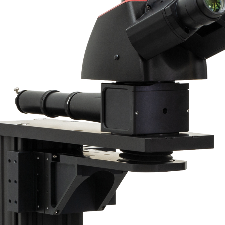
Click to Enlarge
Figure 2.1 This Cerna® microscope kit features a single-cube epi-illuminator module.

Click to Enlarge
Figure 2.2 The front cover of the epi-illuminator module is removed by unscrewing two M2 screws to install a filter cube (not included). Magnets on the cover and the housing ensure that the filter cube is positioned correctly when the door is replaced.
Click to Enlarge
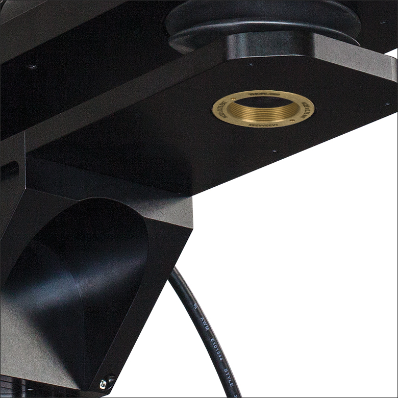
Figure 2.3 The back of the epi-illuminator module includes a removable, SM1-threaded adapter that accepts liquid light guides.
Trans-Illumination (Brightfield Imaging)
Features
- Supports Brightfield Illumination in the Visible and NIR
- Accepts Thorlabs' Illumination Kits (Visible Illumination Kit Included)
- Motorized Condenser Focusing Module with 1" Travel
This Cerna microscope configuration includes both a trans-illumination module and condenser, designed to direct and condition visible and/or IR illumination generated by one of our Illumination Kits into the optical path. Please see the full web presentation for additional information.
Bright visible light is generated by the included illumination kit (Item # WFA1010), which uses one of Thorlabs' Mounted LEDs (Item # MWWHL4). The module features additional ports and a filter cube holder to allow for later expansion with IR or other wavelength LEDs. Please contact Technical Support with inquiries.
Click to Enlarge
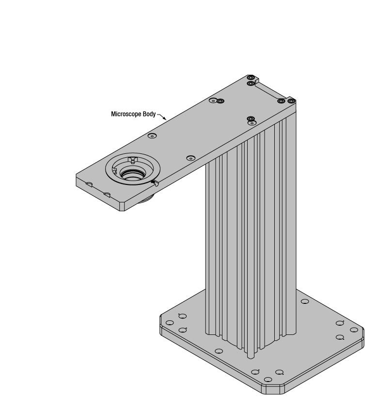
Figure 2.5 The microscope body is based on a 95 mm optical rail.
Microscope Body
Features
- Large Working Volume: Optical Path is 7.74" (196.6 mm) Away from Edge of Rail
- Linear Dovetail Surface Allows Modules to be Added and Removed
- 350 mm Body Height to Accommodate Sample Stages Mounted on Rigid Stands or Fixed Arms
- Motorized Objective Focusing Module with 1" Travel
- Mechanically Compatible with Thorlabs' 95 mm Rail Platforms
The backbone of this Cerna microscope kit is the 350 mm tall microscope body based on Thorlabs' 95 mm Precision Optical Rails, providing stable long-term support and excellent vibrational damping. Its linear dovetail mounting surface allows modules to be removed when they are not needed, freeing additional workspace and opening the door to user customization. For alternate rail heights please see the full web presentation.

Click to Enlarge
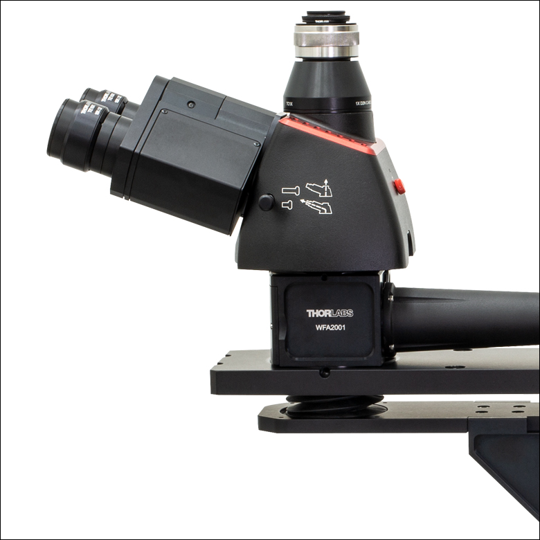
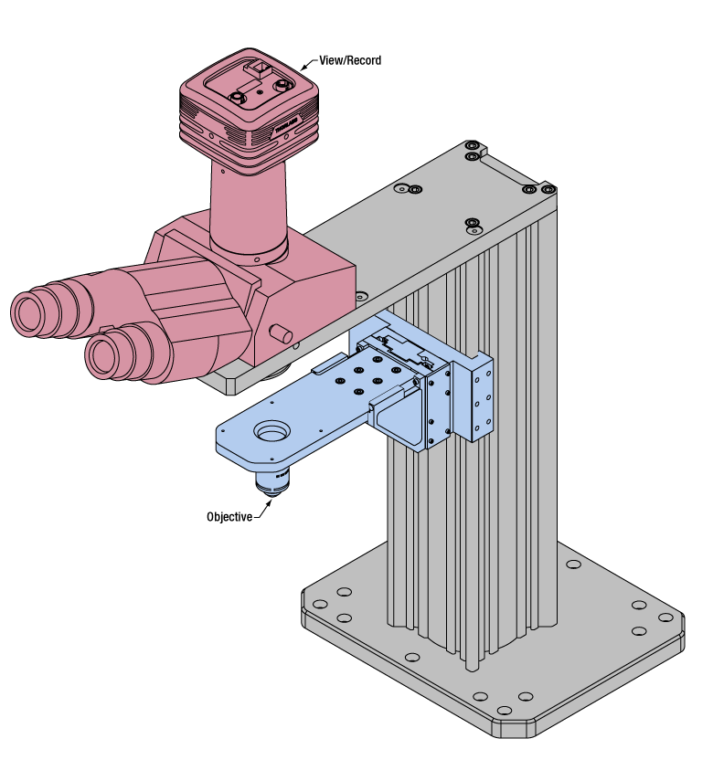
Figure 2.6 This microscope includes trinoculars with a camera port for widefield viewing.
Widefield Viewing
Add-On: Widefield Viewing
Features
- Trinoculars for Viewing Visible Light from the Sample
- Fixed Magnification Camera Port with C-Mount Accepts Most Industry-Standard Cameras
- Trinoculars with 10X Eyepiece Magnification and Adjustable Interpupil Distance
Widefield viewing is provided by trinoculars and a camera tube. The eyepieces feature an adjustable interpupil distance and rotate individually to allow the focus to be coarsely adjusted for each eye.
The included camera tube contains all of the optics needed to image the light from the objective onto a camera sensor. External C-mount (1.00-32") threads on the top of the camera tube accept Thorlabs' scientific cameras, as well as cameras from most major manufacturers. For additional viewing port and camera tube options, please see the full web presentation.
Click to Enlarge
Figure 2.7 This microscope kit has a single objective holder (objective not included).
Objective Holders and Objectives
Add-On: Objectives
Features
- Threaded for M32 x 0.75 Objectives
- Included Adapters:
- M25 x 0.75 Internal Threads
- RMS Internal Threads
The Single-Objective Nosepiece connects to the motorized mounting arm on the microscope body via six M4 counterbores to provide 1" of motorized vertical translation of the objective. The nosepiece features an M32 x 0.75 threaded port for mounting objectives and four 4-40 through taps for 60 mm cage system compatibility. Two objective thread adapters are also included to provide compatibility with other common objective threads: M25 x 0.75 and RMS. Microscope objectives are available for purchase separately from Thorlabs, and we can also order other objectives upon request. To mount multiple objectives, please see the full web presentation for other mounting options. Keep in mind that the total system magnification will depend upon the objective chosen; see the Objective, Scan, and Tube Lens Tutorial for details.
This kit configuration is constructed from our modular Cerna® components. See the comprehensive list below for each included item.
| Item # | Qty. | Description | Photo (Click to Enlarge) |
|---|---|---|---|
| Microscope Body | |||
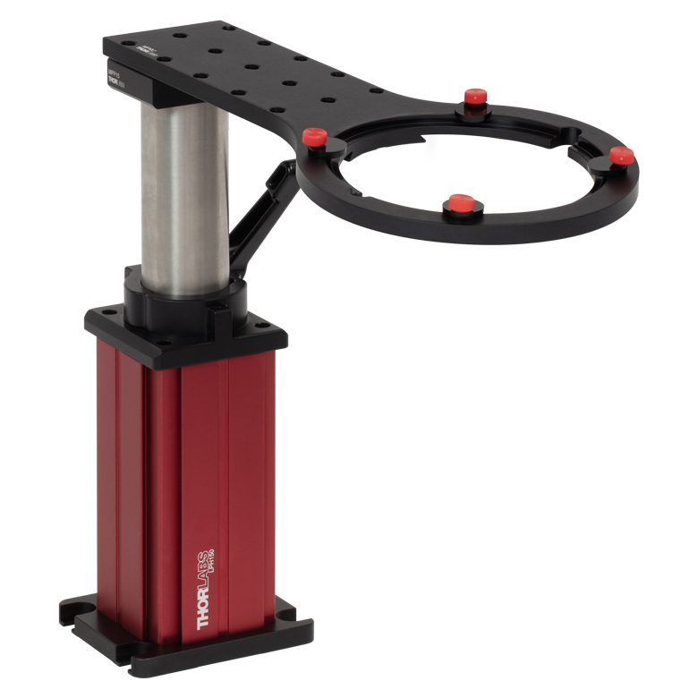
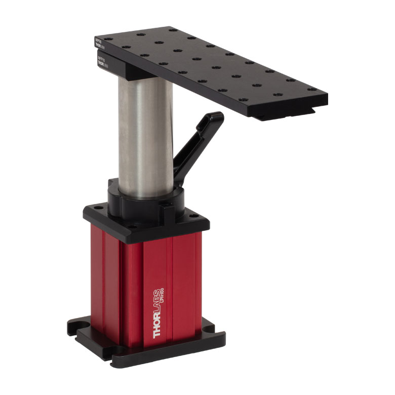
| CEA1350 | 1 | Cerna Microscope Body with Epi-Illumination Arm, 350 mm Tall | 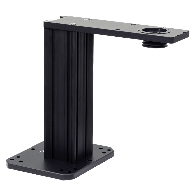 |
| Widefield Viewing | |||
| LAURE1 | 1 | Trinoculars with Eyepieces |  |
| TC1X | 1 | 1X Camera Tube with C-Mount | 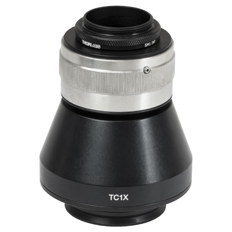 |
| Epi-Illumination | |||
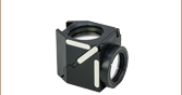
| WFA2001 | 1 | Single-Cube Epi-Illuminator Module (Filter Cube Not Included) |  |
| Condenser | |||
| CSC1001 | 1 | Nikon FN-C LWD Condenser, 0.78 NA | 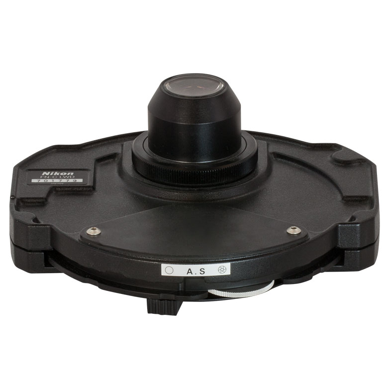 |
| Objective & Condenser Mounts | |||
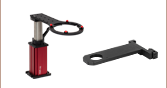
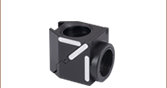
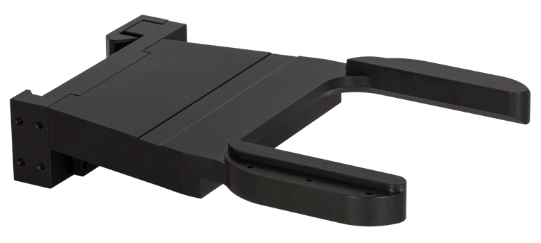
| CSN100 | 1 | Single-Objective Nosepiece | 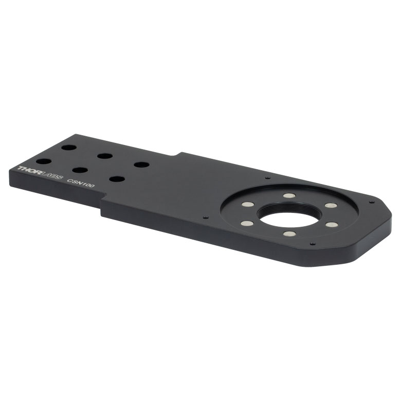 |
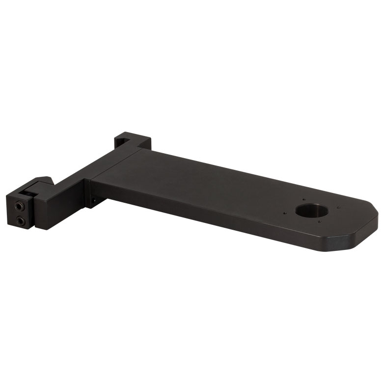
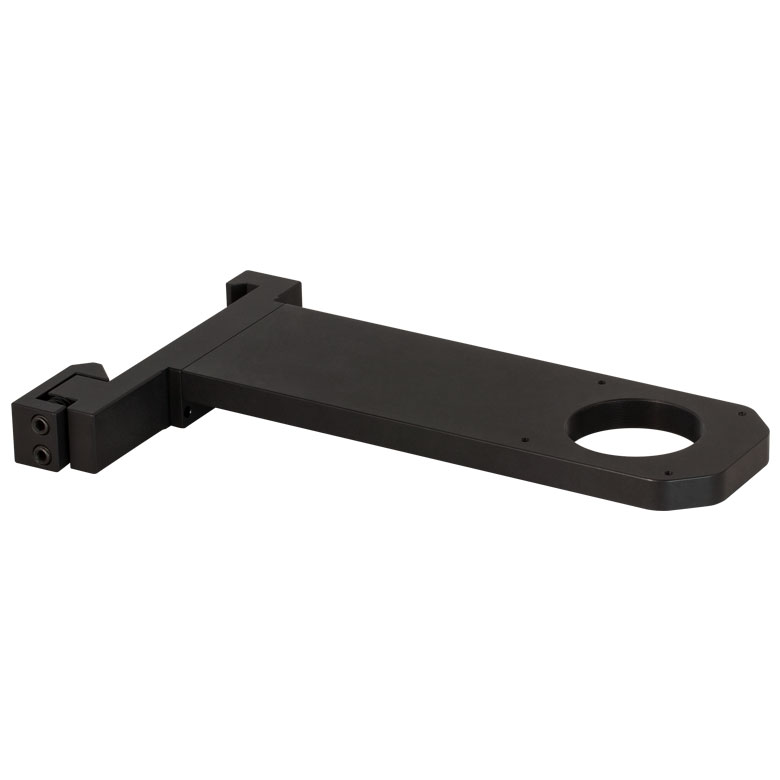
| CSA2000 | 1 | Condenser Mounting Arm with ±2 mm Travel in X and Y | |
| PLSZ | 2 | Motorized Module with 1" Travel, 95 mm Dovetail | 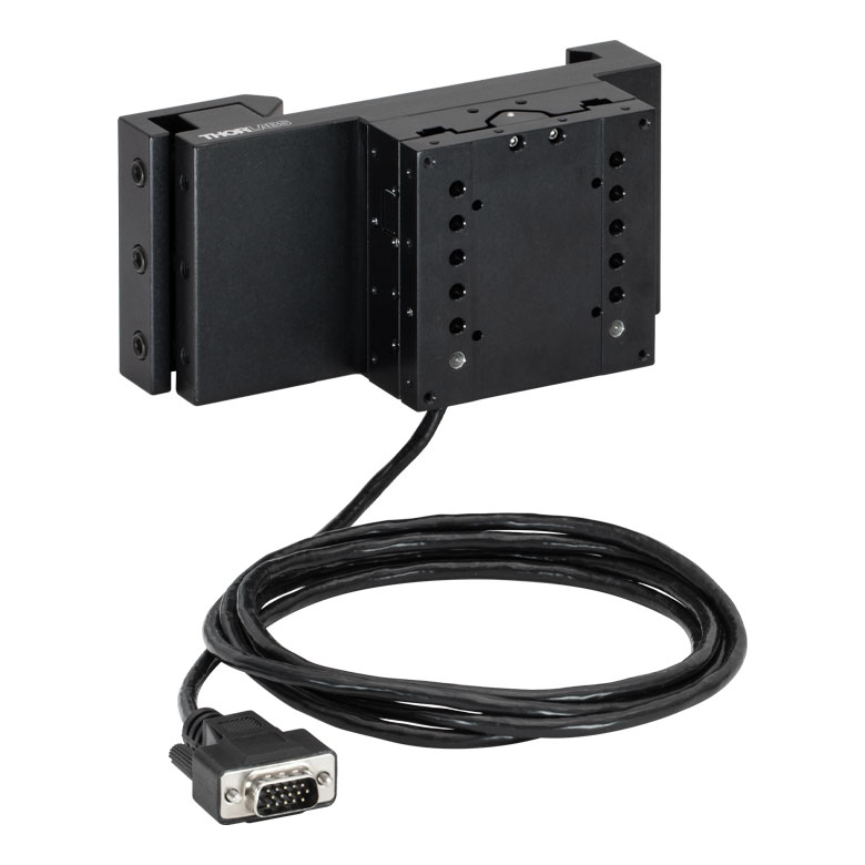 |
| PLSZ1 | 2 | Angle Bracket for Edge-Mounted Arms |  |
| MCMK3 | 1 | 3-Knob USB HID Joystick | 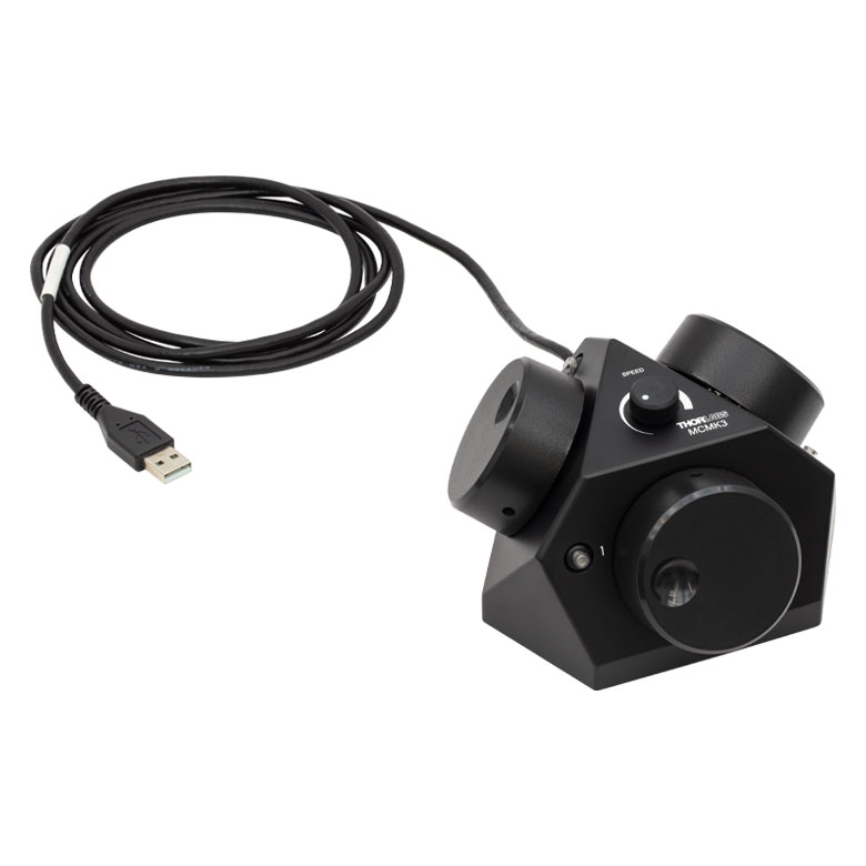 |
| MCM301 | 1 | Three-Channel Controller for Motorized Rigid Stands and PLS Series Stage |  |
| Trans-Illumination | |||
| WFA1000 | 1 | Brightfield Illumination / DIC Imaging Module | 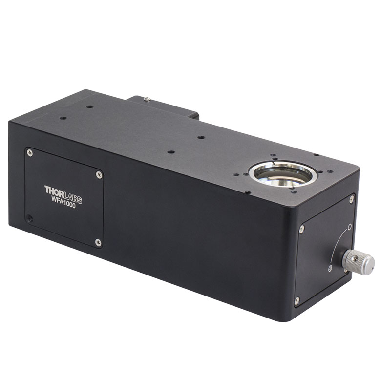 |
| WFA0150 | 1 | Transmitted Light Module Dovetail Clamp | 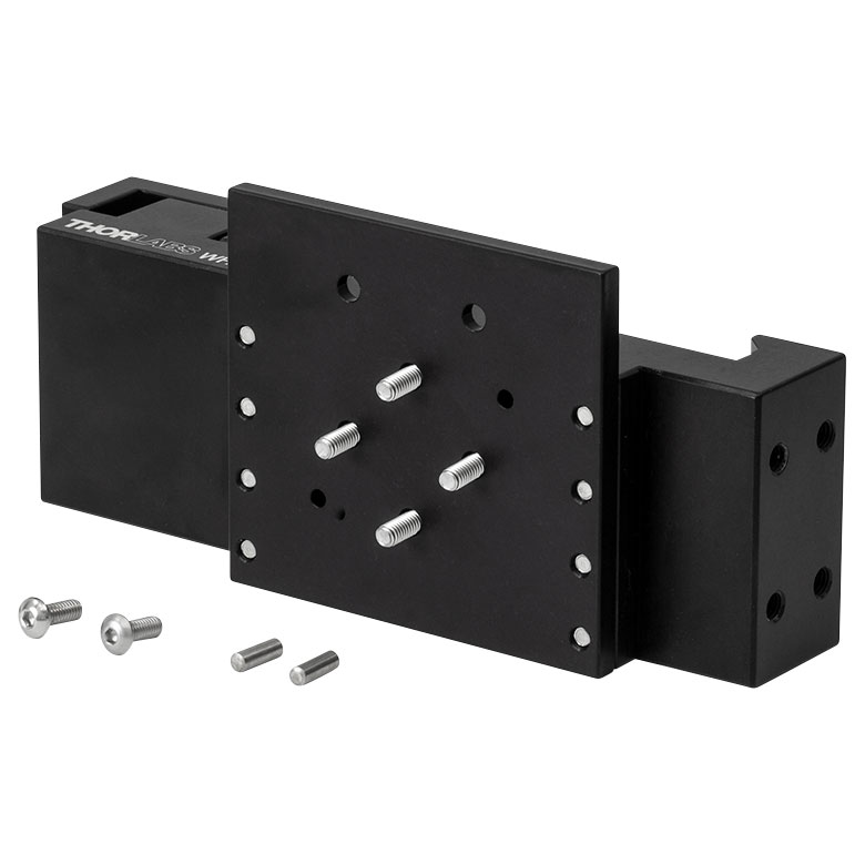 |
| Illumination Kit | |||
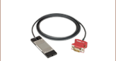
| WFA1010 | 1 | Visible Illumination Kit | 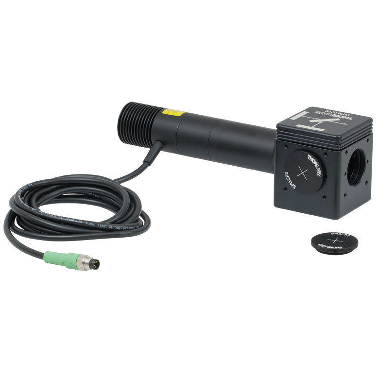 |
| LEDD1B | 1 | T-Cube™ LED Driver, 1200 mA Max Drive Current (Power Supply Not Included) | 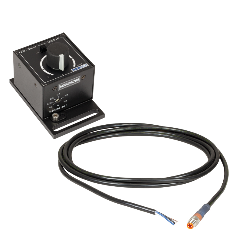 |
| KPS201 | 1 | 15 V Power Supply Unit for a Single K-Cube® or T-Cube™ | 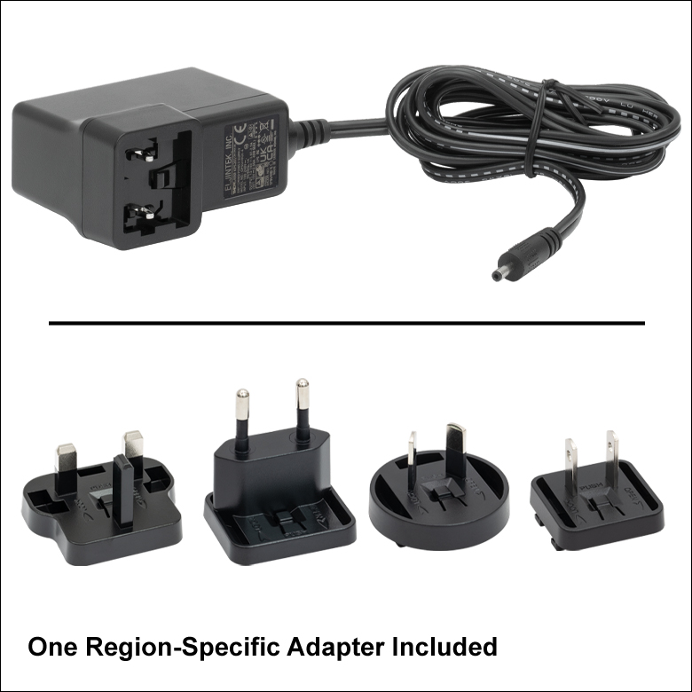 |
| Objective Threading Adapters | |||
| M32M25S | 1 | External M32 x 0.75 Threads and Internal M25 x 0.75 Threads | 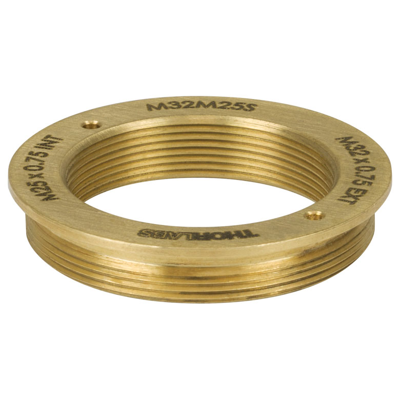 |
| M32RMSS | 1 | External M32 x 0.75 Threads and Internal RMS Threads | 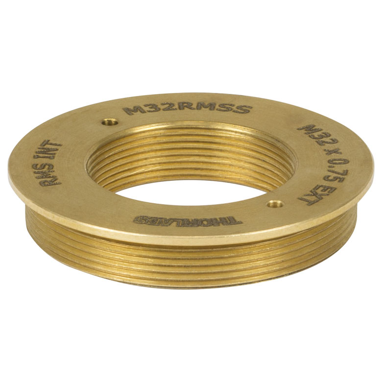 |
Application-Optimized Cerna Microscopes
Developed in collaboration with our colleagues in the field, the Cerna microscopy platform is uniquely modular and flexible, making it adaptable to a wide range of demanding experimental requirements. If you would like to work with our application specialists, engineers, and sales team to design your own microscope, please email ImagingSales@thorlabs.com.
Selected Accessories
In order to image with this microscope, it is necessary to add scientific cameras, an epi-illumination source, filter cubes and filter sets, objectives, and sample holders. It is often possible to improve the quality of your experimental data by carefully selecting accessories that complement your specific experiment. To that end, we have ensured that Cerna® microscopes are compatible with a wide range of accessories. The information below compares the Cerna-compatible components that are manufactured or sold by Thorlabs. We have also indicated when it is possible to use equipment designed by other manufacturers.
Content
- Scientific Cameras for Widefield Viewing
- Illumination Sources for Epi-Illumination
- Filter Cubes and Filter Sets for Epi-Fluorescence
- Objectives
- Sample Holders
Click to Enlarge
Figure 4.1 The camera port provides a fixed magnification for light from the sample.
Scientific Cameras for Widefield Viewing
- Visualize the Field of View at a Computer
- Any C-Mount Camera is Compatible with a Cerna Microscope
Thorlabs offers scientific cameras optimized for a range of imaging needs. Cameras allow the field of view to be displayed on a computer screen and saved for later reference. Viewing your sample from a computer also enables remote sample positioning using our motion control accessories (see below), allowing samples to be moved in sensitive setups without introducing additional vibrations from your hands.
This Cerna microscope configuration includes a camera tube, which provides a fixed magnification at the image plane.
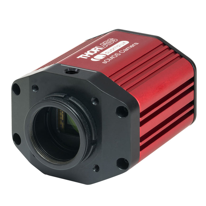
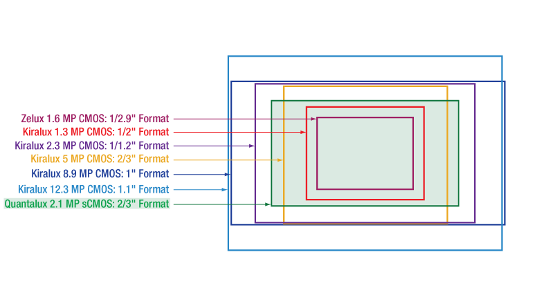
Any camera with C-Mount (1.00"-32) threading is compatible with this microscope. We recommend the CS2100M-USB Quantalux® Scientific sCMOS Camera; see the Table 4.2 for more details. For other options, please see our complete range of scientific cameras.
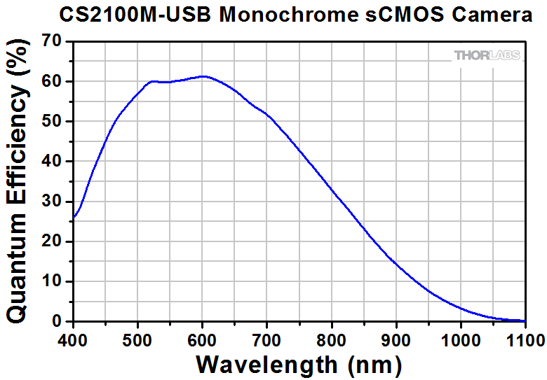
| Table 4.2 CS2100M-USB Specifications | |
|---|---|
| Product Photo (Click to Enlarge) |
 |
| Sensor Type | Monochrome sCMOS |
| Effective Number of Pixels (Horizontal x Vertical) |
1920 x 1080 |
| Imaging Area (Horizontal x Vertical) |
9.6768 mm x 5.4432 mm |
| Pixel Size | 5.04 µm x 5.04 µm |
| Optical Format | 2/3" (11 mm Diagonal) |
| Max Frame Rate | 50 fps (Full Sensor) |
| Sensor Shutter Type | Rolling |
| Peak Quantum Efficiency | 61% at 600 nm |
| PC Interface | USB 3.0 |
| Housing Dimensions | 2.77" x 2.38" x 1.88" (70.4 mm x 60.3 mm x 47.6 mm) |
Illumination Sources for Epi-Illumination

Click to Enlarge
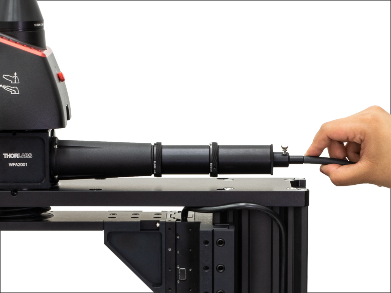
Figure 4.3 A Thorlabs Mounted LED can be threaded into the epi-illuminator module and secured using the included locking ring.
Mounted LEDs
- Long Lifetimes (>10 000 Hours for LEDs Shown Here)
- Output can be Modulated with Suitable Driver
- Integrated EEPROM for Automated Driver Configuration
- Link to Full Web Presentation
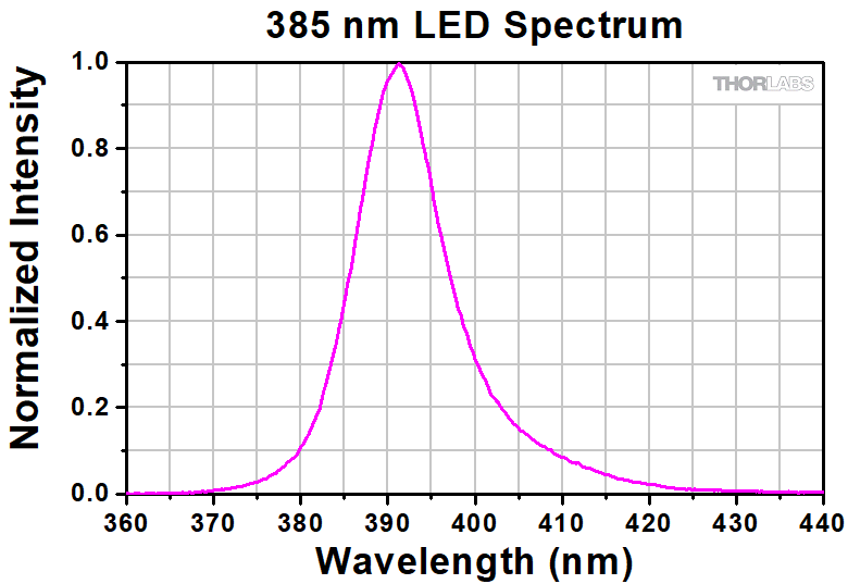
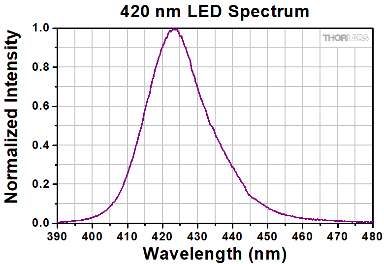
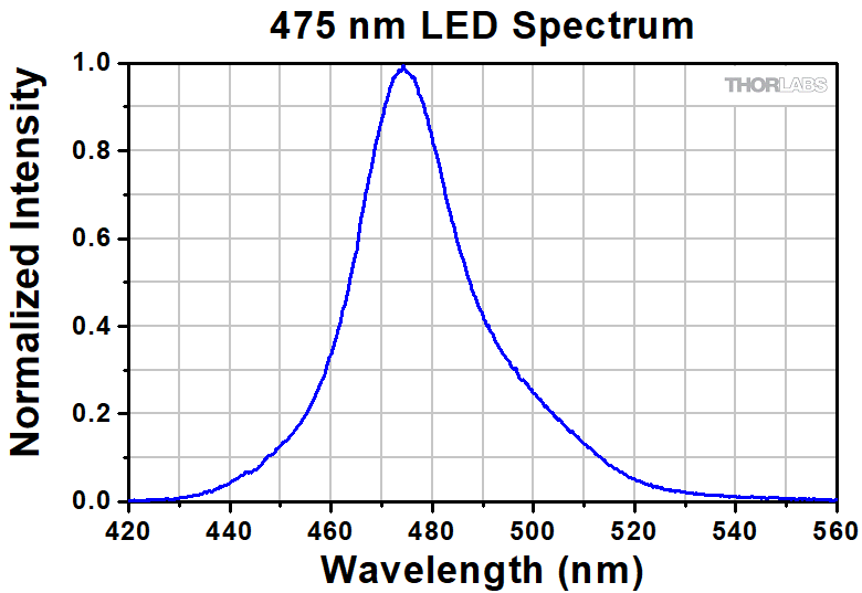
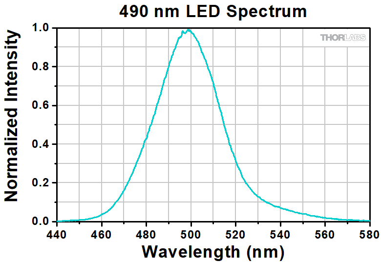
The epi-illuminator module included with this microscope kit is compatible with Thorlabs' LEDs. Selected LEDs that emit at important visible and NIR wavelengths are outlined in Table 4.5. We offer a much wider range of LEDs than the four presented here, at wavelengths from 265 nm to 1650 nm, all of which are compatible with the CM1003 Cerna microscope. For our full selection, please see their full web presentation. Please note that the drivers needed to power the LEDs are sold separately.
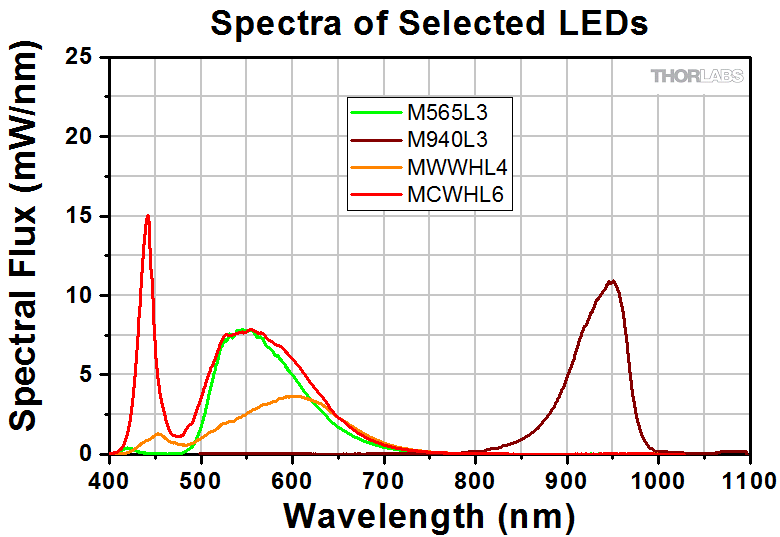
Click to Enlarge
Figure 4.4 This graph compares the spectra of selected Thorlabs LEDs; spectra are scaled for reference.
| Table 4.5 Selected LEDs | |||
|---|---|---|---|
| Item #a | Colorb,c | Output Power (Typical)b | Compatible Drivers |
| M565L3 | Lime Green (565 nm) | 979 mW | LEDD1B DC2200 DC4100 DC4104 |
| M940L3 | IR (940 nm) | 1000 mW | |
| MWWHL4 | Warm White (3000 Kd) | 640 mW | |
| MCWHL7 | Cold White (6500 Kd) | 1370 mW | |
| Full Web Presentation for Mounted LEDs | |||
Click to Enlarge
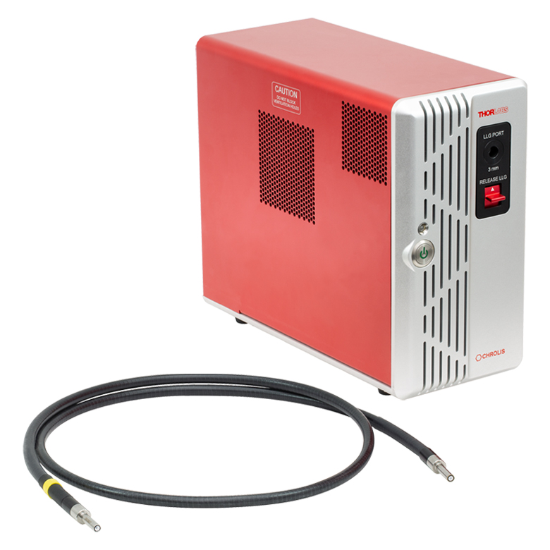
Figure 4.8 Chrolis 6-Wavelength High-Power LED Source with Included LLG03-4H Liquid Light Guide
Click to Enlarge
Figure 4.6 The liquid light guide can be secured by tightening the thumbscrew on the included adapter.
Chrolis™ 6-Wavelength High-Power LED Sources
- Combine 6 User-Changeable, High-Power LEDs into One Liquid Light Guide
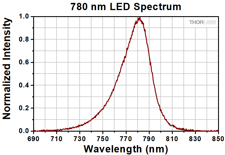
- Eleven Wavelength Options Cover All Known Fluorophores Between 365 nm and 780 nm (See Table 4.7)
- Control LEDs Using Included Software with GUI
- Typical Lifetime of LED Modules is ≥10 000 Hours
- Link to Full Web Presentation
Thorlabs' Chrolis LED sources are user-configurable light engines that efficiently combine the output of six LEDs into a single liquid light guide (LLG). They are ideal for fluorescence imaging that requires up to six wavelengths of light. These sources are available in two pre-set configurations, as well as custom configurations; please see Table 4.7 for the LED options and the full web presentation for more details. As shown in Figure 4.6, the Chrolis sources are compatible with the epi-illuminator module, which includes an adapter that accepts Ø3 mm core liquid light guides.
| Table 4.7 Chrolis LED Options | |||||||||||
|---|---|---|---|---|---|---|---|---|---|---|---|
| Nominal Wavelength |
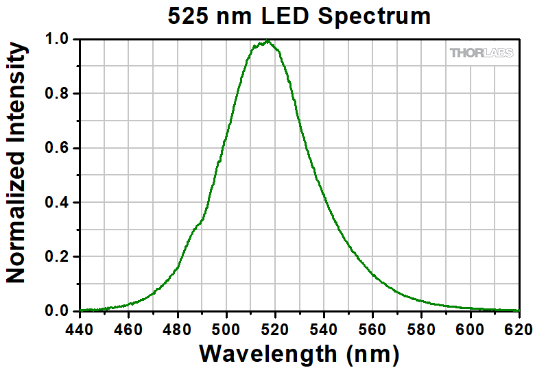
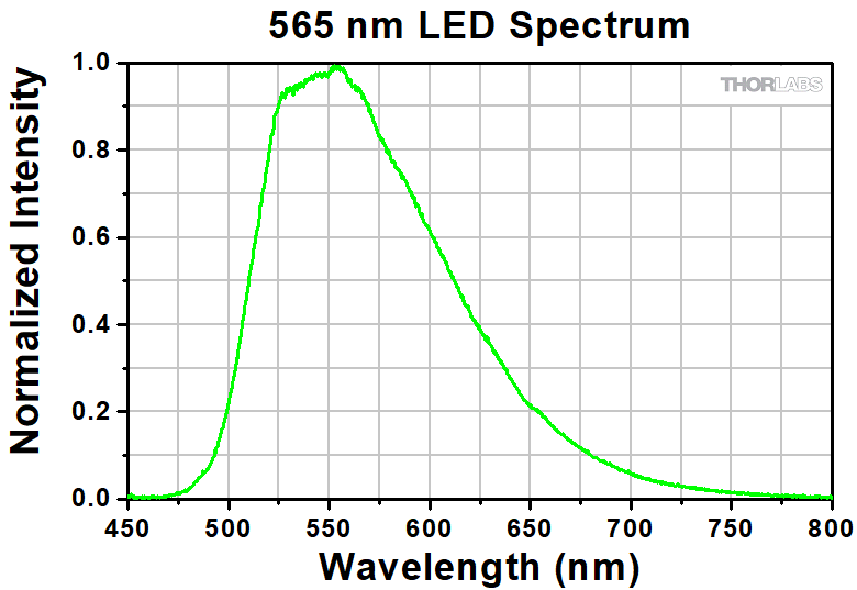
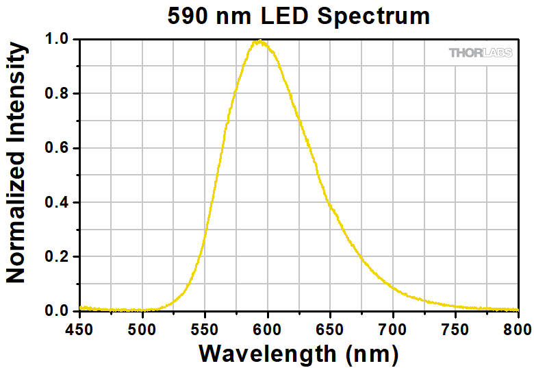
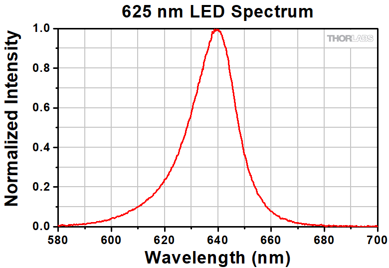
365 nm | 385 nm | 405 nma | 420 nma | 475 nm | 490 nm | 525 nmb | 565 nmb | 590 nm | 625 nm | 780 nm |
| Color (Click for Spectrum) |
UV | UV | UV | Violet | Blue | Blue | Green | Lime | Amber | Red | IR |
| Typical Output Powerc |
1130 mW | 1250 mW | 900 mW | 710 mW | 630 mW | 120 mW | 180 mW | 350 mW | 140 mW | 490 mW | 40 mW |
Filter Cubes and Filter Sets
- Tune Epi-Illumination Source for the Excitation and Detection of Specific Fluorophores
- Easily Mount Filter Sets in the MDFM-MF2
- Each Thorlabs Set Consists of an Excitation Filter, an Emission Filter, and a Dichroic Mirror
- Utilize Fluorescence Filters from Other Major Manufacturers
- Other Filter Sets Available
The epi-illumination module included with this Cerna microscope kit accepts the MDFM-MF2 Filter Cube. This filter cube is designed to hold one Ø25 mm emission filter (up to 5 mm thick), one Ø25 mm excitation filter (up to 3.5 mm thick), and one 25 mm x 36 mm dichroic mirror (up to 1.1 mm thick), as shown in Video 4.10, allowing Cerna microscopes to be compatible with filters from all major manufacturers.
Several popular filter sets are listed with their target fluorophores in Table 4.11. Each set includes an excitation filter, an emission filter, and a dichroic mirror. Please see the full web presentation for the entire line of Thorlabs' filter sets.
|
Video 4.10 Installation of a Filter Set and Filter Cube into
the Single-Cube Epi-Illuminator Module
|
|
|||||||||||||||||||||||||||||||||||||
Objectives
- Cerna Microscope Kit 3 Directly Accepts Objectives with M32 x 0.75 Threads
- Includes Thread Adapters for Compatibility with Objectives from Major Manufacturers
- M25 x 0.75-Threaded Objectives (Nikon)
- RMS-Threaded Objectives (Olympus)
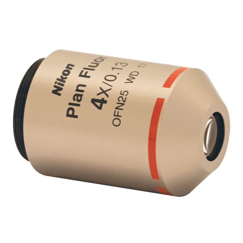
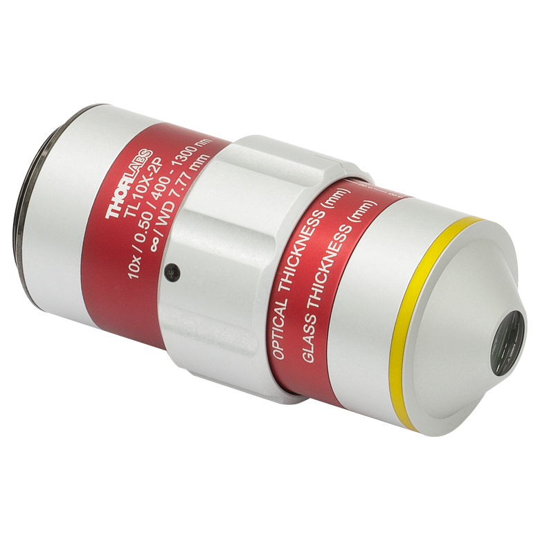
The nosepiece of this microscope has one M32 x 0.75-threaded bore for mounting objectives. The M32 x 0.75 thread standard offers a larger back aperture than previous standards and by newer widefield microscope objectives such as Thorlabs' TL10X-2P and TL15X-2P multiphoton apochromatic microscope objectives. M25 x 0.75- and RMS-threaded adapters are included for compatibility with most objectives from Olympus and Nikon. Shown in Table 4.12 are selected widefield objectives that are commonly used with this microscope kit. They can be mounted in the microscope’s CSN100 Single-Objective Holder directly or by using the M32M25S Brass Microscope Adapter (both included in this configuration). We offer other objectives and can order objectives outside our catalog upon request.
| Table 4.12 Common Widefield Objectives | |||||||
|---|---|---|---|---|---|---|---|
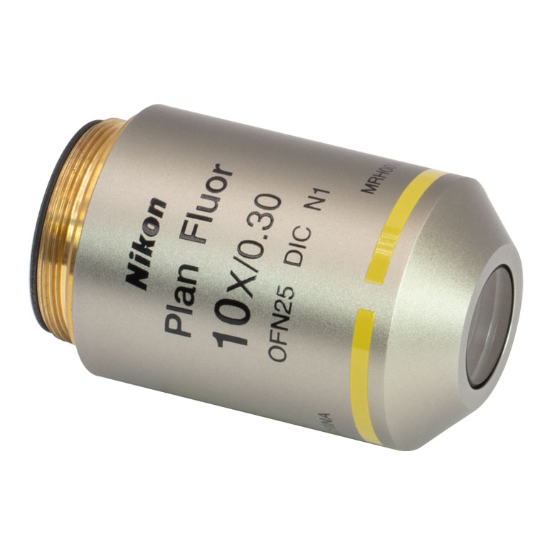
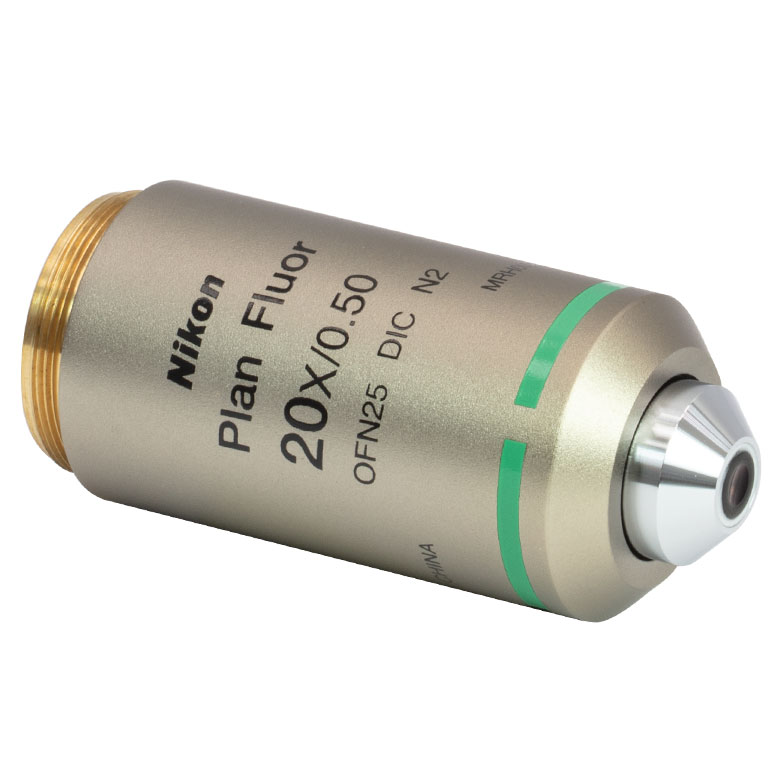
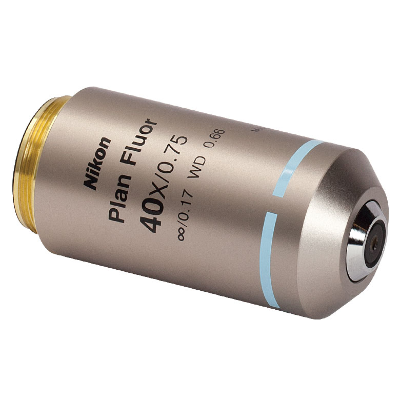
| Item # | N4X-PF | N10X-PF | TL10X-2P | TL15X-2P | N20X-PF | N40X-PF | N60X-PF |
| Photo (Click to Enlarge) |
 |
 |
 |
 |
 |
 |
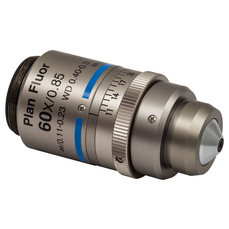 |
| Magnification | 4X | 10X | 10X | 15X | 20X | 40X | 60X |
| Numerical Aperture (NA) | 0.13 | 0.3 | 0.50 | 0.70 | 0.50 | 0.75 | 0.85 |
| Working Distance (WD) | 17.2 mm | 16 mm | 7.77 mm | 2.6 mma | 2.1 mm | 0.66 mm | 0.31 - 0.4 mm |
| Threading | M25 x 0.75 | M32 x 0.75 | M25 x 0.75 | ||||
Click to Enlarge
Figure 4.15 Slide Holder in a Cerna® Microscope

Sample Stages and Holders
- Rigid Stands to Hold Samples Underneath and Around the Objectives
- Designed for Slides, Petri Dishes, Well Plates, Recording Chambers, Micromanipulators, and Custom Inserts
- Translation Stages with 1" of X and Y Travel Available
- Fixed Arms Allow Fast XY Stage, Lens Tubes, and/or Cage Systems to be Placed Directly Into the Optical Path
- CSA1000: For Our MLS203-1 Fast XY Scanning Stage
- CSA1001: For Ø1" Lens Tubes and 30 mm Cage Systems
- CSA1002: For Ø2" Lens Tubes and 60 mm Cage Systems
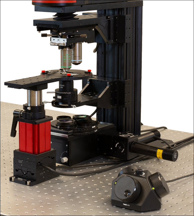
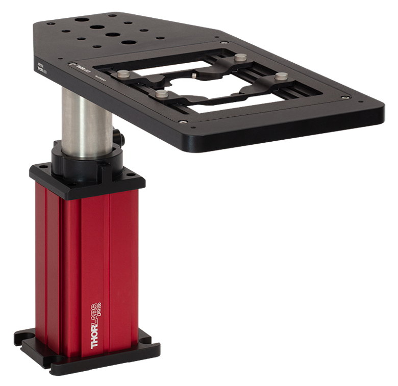
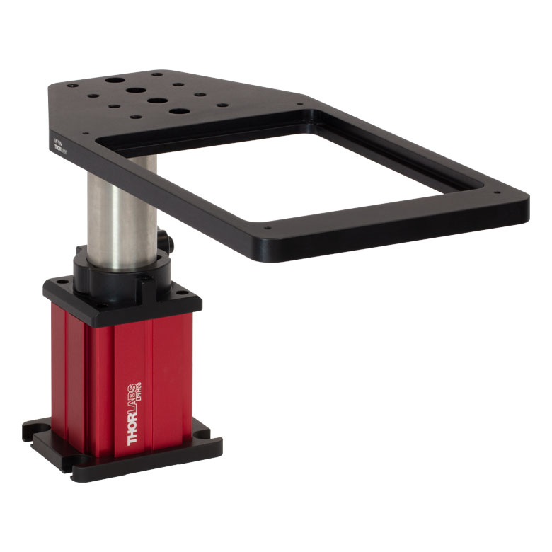
Thorlabs offers highly configurable solutions for mounting your sample beneath the objective of the Cerna Microscope. Rigid stands are available with multiple platform styles that can accept slides, petri dishes, recording chambers, micromanipulators, and custom inserts. The included collar makes them lockable at a height and angle chosen by the user. We also manufacture translation stages for these rigid stands that provide motorized horizontal translation of the sample.
Our fixed arms enable the sample stage to be attached directly to the microscope body via a dovetail that extends the full height of the microscope body, allowing the arms to be positioned anywhere along the body height. For a pre-configured sample holder solution, use the CSA1000 fixed arm with the MLS203-1 Fast XY Scanning Stage. This stage is compatible with our MZS500-E Piezo-Driven Insert, which adds high-resolution Z-axis adjustments. Alternatively, the CSA1001 and CSA1002 rigid arms are compatible with Thorlabs' wide selection of optomechanical components, allowing custom sample holder configurations and additional optics to be easily integrated into this microscope.
Several common options are outlined in Tables 4.16 and 4.17, while our full selection of sample holders can be explored in the Cerna Components presentation.
Table 4.16 Rigid Stands
 Click to Enlarge MP10M(/M) Rigid Stand with Rectangular Insert Holder
|
 Click to Enlarge MPRC(/M) Recording Chamber Holder, MPP15 Post, and
|
 Click to Enlarge MP10(/M) Rigid Stand with Platform
|
Table 4.17 Fixed Arms
CSA1000 Fixed Arm
|
CSA1001 Fixed Arm
|
CSA1002 Fixed Arm
|
| Posted Comments: | |
| No Comments Posted |
Click on the different parts of the microscope to explore their functions.
Elements of a Microscope
This overview was developed to provide a general understanding of a Cerna® microscope. Click on the different portions of the microscope in Figure 77A or use the links below to learn how a Cerna microscope visualizes a sample.
Terminology
Arm: Holds components in the optical path of the microscope.
Bayonet Mount: A form of mechanical attachment with tabs on the male end that fit into L-shaped slots on the female end.
Bellows: A tube with accordion-shaped rubber sides for a flexible, light-tight extension between the microscope body and the objective.
Breadboard: A flat structure with regularly spaced tapped holes for DIY construction.
Dovetail: A form of mechanical attachment for many microscopy components. A linear dovetail allows flexible positioning along one dimension before being locked down, while a circular dovetail secures the component in one position. See the Microscope Dovetails tab or here for details.
Epi-Illumination: Illumination on the same side of the sample as the viewing apparatus. Epi-fluorescence, reflected light, and confocal microscopy are some examples of imaging modalities that utilize epi-illumination.
Filter Cube: A cube that holds filters and other optical elements at the correct orientations for microscopy. For example, filter cubes are essential for fluorescence microscopy and reflected light microscopy.
Köhler Illumination: A method of illumination that utilizes various optical elements to defocus and flatten the intensity of light across the field of view in the sample plane. A condenser and light collimator are necessary for this technique.
Nosepiece: A type of arm used to hold the microscope objective in the optical path of the microscope.
Optical Path: The path light follows through the microscope.
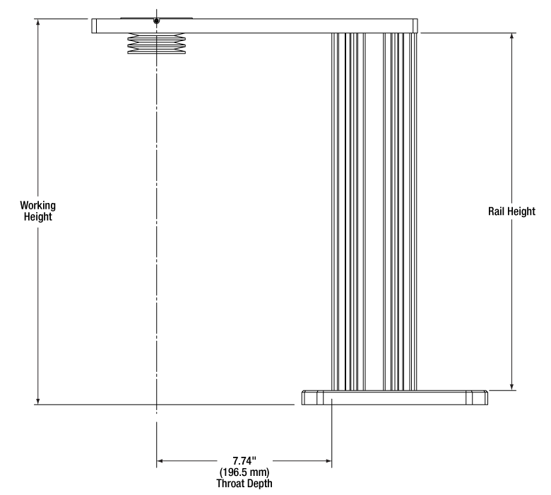
Rail Height: The height of the support rail of the microscope body.
Throat Depth: The distance from the vertical portion of the optical path to the edge of the support rail of the microscope body. The size of the throat depth, along with the working height, determine the working space available for microscopy.
Trans-Illumination: Illumination on the opposite side of the sample as the viewing apparatus. Brightfield, differential interference contrast (DIC), Dodt gradient contrast, and darkfield microscopy are some examples of imaging modalities that utilize trans-illumination.
Working Height: The height of the support rail of the microscope body plus the height of the base. The size of the working height, along with the throat depth, determine the working space available for microscopy.
 Click to Enlarge
Figure 77C Cerna Microscope Body
Click to Enlarge
Figure 77B Body Details
Microscope Body
The microscope body provides the foundation of any Cerna microscope. The support rail utilizes 95 mm rails machined to a high angular tolerance to ensure an aligned optical path and perpendicularity with the optical table. The support rail height chosen (350 - 600 mm) determines the vertical range available for experiments and microscopy components. The 7.74" throat depth, or distance from the optical path to the support rail, provides a large working space for experiments. Components attach to the body by way of either a linear dovetail on the support rail, or a circular dovetail on the epi-illumination arm (on certain models). Please see the Microscope Dovetails tab or here for further details.
 Click to Enlarge
Figure 77D Illumination with a Cerna microscope can come from above (yellow) or below (orange). Illumination sources (green) attach to either.
Illumination
Using the Cerna microscope body, a sample can be illuminated in two directions: from above (epi-illumination, see yellow components in Figure 77D) or from below (trans-illumination, see orange components in Figure 77D).
Epi-illumination illuminates on the same side of the sample as the viewing apparatus; therefore, the light from the illumination source (green) and the light from the sample plane share a portion of the optical path. It is used in fluorescence, confocal, and reflected light microscopy. Epi-illumination modules, which direct and condition light along the optical path, are attached to the epi-illumination arm of the microscope body via a circular D1N dovetail (see the Microscope Dovetails tab or here for details). Multiple epi-illumination modules are available, as well as breadboard tops, which have regularly spaced tapped holes for custom designs.
Trans-illumination illuminates from the opposite side of the sample as the viewing apparatus. Example imaging modalities include brightfield, differential interference contrast (DIC), Dodt gradient contrast, oblique, and darkfield microscopy. Trans-illumination modules, which condition light (on certain models) and direct it along the optical path, are attached to the support rail of the microscope body via a linear dovetail (see Microscope Dovetails tab or here). Please note that certain imaging modalities will require additional optics to alter the properties of the beam; these optics may be easily incorporated in the optical path via lens tubes and cage systems. In addition, Thorlabs offers condensers, which reshape input collimated light to help create optimal Köhler illumination. These attach to a mounting arm, which holds the condenser at the throat depth, or the distance from the optical path to the support rail. The arm attaches to a focusing module, used for aligning the condenser with respect to the sample and trans-illumination module.
 |
 |
 |
 |
 |
 |
 |
 |
| Epi-Illumination Modules | Breadboards & Body Attachments |
Brightfield | DIC | Dodt | Condensers | Condenser Mounting | Light Sources |
 Click to Enlarge
Figure 77E Light from the sample plane is collected through an objective (blue) and viewed using trinocs or other optical ports (pink).
Sample Viewing/Recording
Once illuminated, examining a sample with a microscope requires both focusing on the sample plane (see blue components in Figure 77E) and visualizing the resulting image (see pink components).
A microscope objective collects and magnifies light from the sample plane for imaging. On the Cerna microscope, the objective is threaded onto a nosepiece, which holds the objective at the throat depth, or the distance from the optical path to the support rail of the microscope body. This nosepiece is secured to a motorized focusing module, used for focusing the objective as well as for moving it out of the way for sample handling. To ensure a light-tight path from the objective, the microscope body comes with a bellows (not pictured).
Various modules are available for sample viewing and data collection. Trinoculars have three points of vision to view the sample directly as well as with a camera. Double camera ports redirect or split the optical path among two viewing channels. Camera tubes increase or decrease the image magnification. For data collection, Thorlabs offers both cameras and photomultiplier tubes (PMTs), the latter being necessary to detect fluorescence signals for confocal microscopy. Breadboard tops provide functionality for custom-designed data collection setups. Modules are attached to the microscope body via a circular dovetail (see the Microscope Dovetails tab or here for details).


 Click to Enlarge
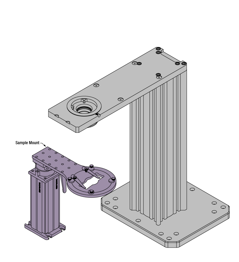
Figure 77F The rigid stand (purple) pictured is one of various sample mounting options available.
Sample/Experiment Mounting
Various sample and equipment mounting options are available to take advantage of the large working space of this microscope system. Large samples and ancillary equipment can be mounted via mounting platforms, which fit around the microscope body and utilize a breadboard design with regularly spaced tapped through holes. Small samples can be mounted on rigid stands (for example, see the purple component in Figure 77F), which have holders for different methods of sample preparation and data collection, such as slides, well plates, and petri dishes. For more traditional sample mounting, slides can also be mounted directly onto the microscope body via a manual XY stage. The rigid stands can translate by way of motorized stages (sold separately), while the mounting platforms contain built-in mechanics for motorized or manual translation. Rigid stands can also be mounted on top of the mounting platforms for independent and synchronized movement of multiple instruments, if you are interested in performing experiments simultaneously during microscopy.
 |
 |
 |
 |
 |
| Translating Platforms | Rigid Stands | Translation Stages for Rigid Stands | Motorized XY Stages | Manual XY Stage |
For sample viewing, Thorlabs offers trinoculars, double camera ports, and camera tubes. Light from the sample plane can be collected via cameras, photomultiplier tubes (PMTs), or custom setups using breadboard tops. Click here for additional information about viewing samples with a Cerna microscope.
| Product Families & Web Presentations | |||
 |
|
 |
 |
| Sample Viewing | Breadboards & Body Attachments |
Cameras | PMTs |
Microscope objectives are held in the optical path of the microscope via a nosepiece. Click here for additional information about viewing a sample with a Cerna microscope.
| Product Families & Web Presentations | ||||
 |
 |
 |
 |
 |
| Objectives | Objective Thread Adapters | Parfocal Length Extender | Piezo Objective Scanner | Objective Mounting |
Large and small experiment mounting options are available to take advantage of the large working space of this microscope. Click here for additional information about mounting a sample for microscopy.
| Product Families & Web Presentations | ||||
|
|
|
|
 |
| Translating Platforms | Rigid Stands | Translation Stages for Rigid Stands | Motorized XY Stages | Manual XY Stage |
Thorlabs offers various light sources for epi- and trans-illumination. Please see the full web presentation of each to determine its functionality within the Cerna microscopy platform.
| Product Families & Web Presentations | ||||
 |
 |
 |
 |
|
| Trans-Illumination Kits | Solis™ High-Power LEDs | Mounted LEDs | X-Cite® Lamps | Other Light Sources |
Epi-illumination illuminates the sample on the same side as the viewing apparatus. Example imaging modalities include fluorescence, confocal, and reflected light microscopy. Click here for additional information on epi-illumination with Cerna.
| Product Families & Web Presentations | ||
|
|
|
| Epi-Illumination | Body Attachments | Light Sources |
Trans-illumination illuminates from the opposite side of the sample as the viewing apparatus. Example imaging modalities include brightfield, differential interference contrast (DIC), Dodt gradient contrast, oblique, and darkfield microscopy. Click here for additional information on trans-illumination with Cerna.
| Product Families & Web Presentations | ||||||
|
|
|
|
 |
|
|
| Brightfield | DIC | Dodt | Condensers | Condenser Mounting | Illumination Kits | Other Light Sources |
The microscope body provides the foundation of any Cerna microscope. The 7.74" throat depth provides a large working space for experiments. Click here for additional information about the Cerna microscope body.
| Product Families & Web Presentations | |
 |
 |
| Microscope Bodies | Microscope Translator |
This microscope configuration can be tailored to your particular imaging needs through the use of our kit functionality. Its components can be added all at once to the shopping cart using the "Add Kit" button at the bottom of the ordering area, or individually using the shopping cart icon next to each item. Items may be removed from the default item list by changing the value in the "Qty" box to 0 before clicking the "Add Kit" button. Once added, peruse our catalog of modular microscope components to further customize the microscope kit in your cart. A discount is offered when a sufficient number of components are purchased. Please see the Kit Components tab for additional information about each component in this microscope kit.