 Products Home / Imaging Systems / OCT Imaging Systems & Components / Application Articles / In Utero Imaging of Mouse Development
In Utero Imaging of Mouse Development

Please Wait
High-Resolution Imaging of In Utero Mouse Development Using OCT
Featured Researchers:
S. H. Syed, K. V. Larin, M. E. Dickinson, and I. V. Larina
Mice are frequently used as a model to study the development and diseases of different human organ systems. The traditional method for studying embryonic mutant phenotypes has been analysis of thin slices of tissue, which requires sacrificing many litters at various stages to determine changes in observable characteristics.
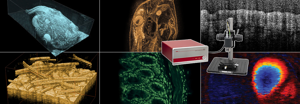
Several in utero methods can provide dynamic information about mouse embryonic development, such as ultrasound, micro-MRI, or microcomputed tomography, but all suffer from a low resolution of 25 – 100 μm, sensitivity to motion, and/or long acquisition times. In contrast, Optical Coherence Tomography (OCT) provides 3D imaging with resolution ranging from 2 – 10 μm with imaging depths of 1 – 3 mm over short acquisition times.
In addition, OCT allows for repetitive, live embryonic imaging in utero from 12.5 days post coitus (dpc), when the decidua surrounding the embryo thins to allow for optical imaging, through 18.5 dpc, which is just prior to birth. In utero OCT measurements can provide dynamic information about embryonic development, allow high-throughput analysis, and eliminate the need for sacrificing multiple litters.
In recently published results, Syed et al. demonstrate the capabilities of OCT to image mouse embryonic development in utero. In the experiment, a portion of the uterus was externalized through a 1 – 2 cm abdominal incision in order to identify and optimize imaging of each embryo. During the procedure, the pregnant female was anesthetized and kept on a heating pad to maintain body temperature.
Once the uterus was externalized, the embryo was imaged through the uterine wall using an OCT system based on Thorlabs’ Swept Source Engine (central wavelength: 1325 nm, bandwidth: 110 nm). Each 3D data set is a reconstruction of a series of 512 x 512 A-Scans taken over a 10 mm x 10 mm x 2.2 mm volume, and each set took around 20 seconds to complete. The resolution of this system was measured as 8 μm in both the lateral and depth directions. The embryos remained alive throughout the imaging sessions, as evidenced by blood flow observed by Doppler OCT measurements.
Figure 1.1 shows 3D reconstructions of the embryonic forelimb at various stages of development between 12.5 and 18.5 dpc using the OCT system. As a comparison, the inset figures show scanning electron micrograph (SEM) images from the Atlas of Mouse Development. The differences in developmental stages are clearly visible, and the OCT images are comparable with those from SEM. For example, the development of individual digits is visible between 12.5 and 13.5 dpc, while the development of nails is visible at 15.5 dpc. Since several mouse models of limb abnormalities associated with human defects have been developed, live OCT imaging can help to further our understanding of these abnormalities in both mice and humans.
OCT is a promising technique for live imaging of mouse embryos and yields dynamic images where structures are easily recognizable. This will significantly advance observational technology needed to screen for mutations and toxins that disrupt normal development.

Figure 1.1 3D reconstructions of an embryonic mouse forelimb at different stages of development imaged in utero using OCT. Insets show scanning electron micrographs at the same developmental stage.
References:
S. H. Syed, K. V. Larin, M. E. Dickinson, and I. V. Larina. Optical coherence tomography for high-resolution imaging of mouse development in utero. J. Biomed. Opt. 16, 046004 (2011).
All mouse manipulation procedures described here were approved by the Animal Care and Use Committee at the University of Houston.
| Posted Comments: | |
| No Comments Posted |